
Page 24 - Zoo Animal Learning and Training
P. 24
Chapter 2: Orthopedic Implants in Neurosurgery 15
screw) has been common practice in veterinary surgery but has
been shown to be of limited value, both in osteoporotic long bones
and in vertebrae [20,21]. If the screw cannot be reoriented to engage
undamaged bone, filling the stripped hole with PMMA prior to
reinserting the screw has been used successfully for the revision of
human pedicle screws and for salvage of stripped holes in feline
radii [22,23].
Types of Screw
Cancellous Screws
Cancellous screws are designed to offer maximum pull‐out resist-
ance in soft and cancellous bones and therefore have a large outer
diameter, a larger pitch, and a smaller core. However, because of
their smaller core, they provide reduced resistance in bending and
shear and may not be appropriate for use in angle‐stable constructs
(such as a locking plate or screws and PMMA internal fixator)
[15,16]. With the exception of self‐drilling screws, a pilot hole
equivalent to, or slightly smaller than, the core diameter of the
screw must be drilled into the bone before inserting the screw.
Tapping the bone to create the bone thread has been shown to Figure 2.7 Detail of the tips of 3.5‐mm regular (top) and self‐tapping
decrease pull‐out strength of cancellous screws and therefore this (bottom) cortical screws showing the cutting flute at the extremity of the
step is often avoided in cancellous bone [24]. However, it may be self‐tapping screw (arrowhead).
useful to get the thread started in the cis‐cortex by turning the tap
for a couple of turns before inserting the screw. If tapping is not
performed, it must be noted that if the tip of the screw contacts a Self‐drilling Screws
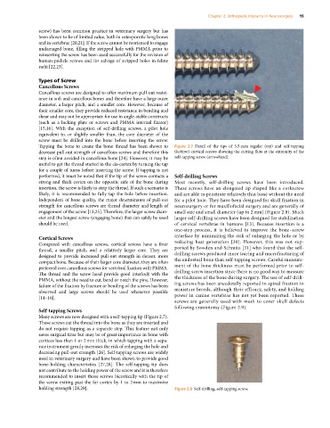
strong and thick cortex on the opposite side of the bone during Most recently, self‐drilling screws have been introduced.
insertion, the screw is likely to strip the thread. If such a scenario is These screws have an elongated tip shaped like a corkscrew
likely, it is recommended to fully tap the hole before insertion. and are able to penetrate relatively thin bone without the need
Independent of bone quality, the major determinants of pull‐out for a pilot hole. They have been designed for skull fixation in
strength for cancellous screws are thread diameter and length of neurosurgery or for maxillofacial surgery and are generally of
engagement of the screw [13,25]. Therefore, the larger screw diam- small size and small diameter (up to 2 mm) (Figure 2.8). Much
eter and the longest screw (engaging bone) that can safely be used larger self‐drilling screws have been designed for stabilization
should be used. of cervical vertebrae in humans [13]. Because insertion is a
one‐step process, it is believed to improve the bone–screw
Cortical Screws interface by minimizing the risk of enlarging the hole or by
Compared with cancellous screws, cortical screws have a finer reducing heat generation [30]. However, this was not sup-
thread, a smaller pitch, and a relatively larger core. They are ported by Sowden and Schmitz [31] who found that the self‐
designed to provide increased pull‐out strength in denser, more drilling screws produced more tearing and microfracturing of
compact bone. Because of their larger core diameter, they are often the endosteal bone than self‐tapping screws. Careful measure-
preferred over cancellous screws for vertebral fixation with PMMA. ment of the bone thickness must be performed prior to self‐
The thread and the screw head provide good interlock with the drilling screw insertion since there is no good way to measure
PMMA, without the need to cut, bend or notch the pins. However, the thickness of the bone during surgery. The use of self‐drill-
failure of the fixation by fracture or bending of the screws has been ing screws has been anecdotally reported in spinal fixation in
observed and large screws should be used whenever possible miniature breeds, although their efficacy, safety, and holding
[14–16]. power in canine vertebrae has not yet been reported. These
screws are generally used with mesh to cover skull defects
following craniotomy (Figure 2.9).
Self‐tapping Screws
Many screws are now designed with a self‐tapping tip (Figure 2.7).
These screws cut the thread into the bone as they are inserted and
do not require tapping as a separate step. This feature not only
saves surgical time but may be of great importance in bone with
cortices less than 1 or 2 mm thick, in which tapping with a sepa-
rate instrument greatly increases the risk of enlarging the hole and
decreasing pull‐out strength [26]. Self‐tapping screws are widely
used in veterinary surgery and have been shown to provide good
bone‐holding characteristics [27,28]. The self‐tapping tip does
not contribute to the holding power of the screw and it is therefore
recommended to insert those screws bicortically with the tip of
the screw exiting past the far cortex by 1 to 2 mm to maximize
holding strength [28,29]. Figure 2.8 Self‐drilling, self‐tapping screw.