
Page 97 - Zoo Animal Learning and Training
P. 97
94 Section I: Diagnostics and Planning
should feel the multitude of perineurium bundles passing
between the tip of the index finger and nail of the thumb. It is
critical to separate the associated artery and vein from the nerve.
This not only decreases morbidity by leaving the vasculature
intact, but also ensures a diagnostic sample is taken and not
a sample from only some of the surrounding connective tissue.
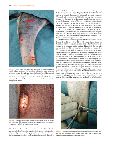
To start, a hemostat is used to separate the nerve, artery, and vein
bundle from surrounding tissue in a 360° dissection. Next, hemo-
stats are used to separate the nerve from the rest of the bundle.
This can be performed by pushing on the edge of the nerve with
the closed tips of hemostats and with downward pressure open-
ing the hemostats to strip away fascia and connective tissue
(Figure 9.3). Once the bundle is identified and completely iso-
lated, a fascicular biopsy is made [5].
The tibial nerve is split in a proximal to distal direction for the
length of the biopsy with a #11 or #15 scalpel blade. It may be
beneficial to have a second scalpel handle or other structure under
the nerve to provide a cutting surface (Figure 9.4). The nerve is
split so that one‐third of its width is taken for the biopsy [5].
Thumb forceps are used to grasp the nerve biopsy at its most
proximal dissection (Figure 9.5). This is the only time the nerve
will be grasped in order to minimize trauma to the biopsy speci-
men. An alternative to manipulating the nerve with forceps is to
suture one‐third of the width of the nerve at the most proximal
aspect of dissection leaving a suture tag to hold. Then the proxi-
mal end of the biopsy is always severed first in a transverse man-
ner followed by the distal aspect (Figure 9.6). This is to minimize
patient discomfort as, even in a surgical plane of anesthesia, sec-
Figure 9.1 Pelvic limb (lateral distal femur and tibia) shown draped for tioning of an intact nerve will arouse the patient because of intense
muscle and nerve biopsies. The dotted line indicates the lateral saphenous pain. The sample is then either pinned or tied to a previously pre-
vein, to be avoided during biopsy of the tibial nerve. The solid arrow indi- pared piece of tongue depressor or tied to the wooden end of a
cates the incision for biopsy of the biceps femoris and gastrocnemius mus- cotton‐tipped applicator. This ensures the nerve will not contract
cles as well as common peroneal nerve. The dashed arrow indicates incision during shipment. The entire sample is placed in 10% formalin.
for biopsy of cranial tibial muscle and double arrow indicates incision for
biopsy of tibial nerve.
Figure 9.2 A biopsy of the cranial tibial muscle has been taken, an absorb-
able hemostatic sponge (arrow) has been placed in the defect, and the fascia
closed using a simple continuous suture pattern.
identification to “strum” the nerve between the tip of the index fin-
ger and nail of the thumb through the sterile gloves. Having initially Figure 9.3 A pair of hemostats has been placed under the tibial nerve bun-
clipped and prepared in a hanging‐leg fashion makes it easier to use dle after 360° dissection and a second pair is being used to dissect away
this strumming technique while maintaining a sterile field. One surrounding vasculature and connective tissue.