
Page 1107 - Equine Clinical Medicine, Surgery and Reproduction, 2nd Edition
P. 1107
1082 CHAPTER 10
VetBooks.ir 10.31 10.32
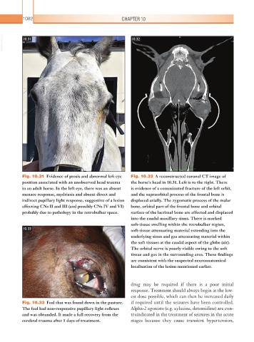
Fig. 10.31 Evidence of ptosis and abnormal left eye Fig. 10.32 A reconstructed coronal CT image of
position associated with an unobserved head trauma the horse’s head in 10.31. Left is to the right. There
in an adult horse. In the left eye, there was an absent is evidence of a comminuted fracture of the left orbit,
menace response, mydriasis and absent direct and and the supraorbital process of the frontal bone is
indirect pupillary light response, suggestive of a lesion displaced axially. The zygomatic process of the malar
affecting CNs II and III (and possibly CNs IV and VI) bone, orbital part of the frontal bone and orbital
probably due to pathology in the retrobulbar space. surface of the lacrimal bone are affected and displaced
into the caudal maxillary sinus. There is marked
soft-tissue swelling within the retrobulbar region,
10.33 soft-tissue attenuating material extending into the
underlying sinus and gas attenuating material within
the soft tissues at the caudal aspect of the globe (air).
The orbital nerve is poorly visible owing to the soft
tissue and gas in the surrounding area. These findings
are consistent with the suspected neuroanatomical
localisation of the lesion mentioned earlier.
drug may be required if there is a poor initial
response. Treatment should always begin at the low-
est dose possible, which can then be increased daily
Fig. 10.33 Foal that was found down in the pasture. if required until the seizures have been controlled.
The foal had non-responsive pupillary light reflexes Alpha-2 agonists (e.g. xylazine, detomidine) are con-
and was obtunded. It made a full recovery from the traindicated in the treatment of seizures in the acute
cerebral trauma after 5 days of treatment. stages because they cause transient hypertension,