
Page 1109 - Equine Clinical Medicine, Surgery and Reproduction, 2nd Edition
P. 1109
1084 CHAPTER 10
VetBooks.ir and cost-effective therapy and could be considered 10.34
in horses with acute brain injury, given via intrave-
nous infusion (MgSO at a rate of 50 mg/kg [approx-
4
imately 250 µmol/kg]). This dose (25 g in a 500-kg
horse) can be easily administered with the first 5–10
litres of intravenous fluid.
Prognosis
The prognosis for acute head trauma to the frontal/
parietal area is highly variable. Recumbency is a poor
prognostic indicator. Ideally, stabilisation of neu-
rological abnormalities should occur within 24–48
hours, with gradual improvement over the next week Fig. 10.34 Horse with a basisphenoid fracture.
and slower improvement from then on. Once a pla-
teau in improvement is encountered, there will usu-
ally be minimal further recovery. The development 10.35
of a midbrain syndrome warrants a poor prognosis,
whereas an uncomplicated cerebral syndrome usu-
ally has a good prognosis, as response to treatment
for brain swelling can be good.
POLL IMPACT
A characteristic pattern of brain injury is observed
when the horse rears over backwards and the back of
the skull hits an overhead structure or the ground.
There is a huge amount of force exerted on the base
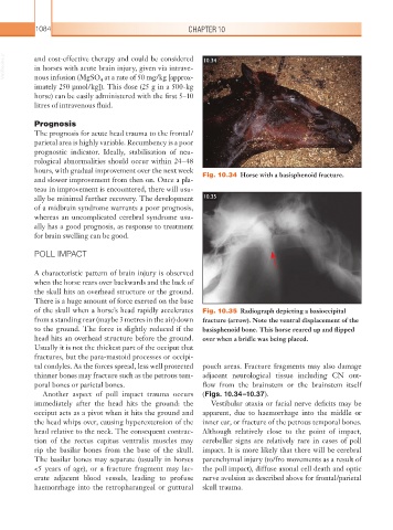
of the skull when a horse’s head rapidly accelerates Fig. 10.35 Radiograph depicting a basioccipital
from a standing rear (maybe 3 metres in the air) down fracture (arrow). Note the ventral displacement of the
to the ground. The force is slightly reduced if the basisphenoid bone. This horse reared up and flipped
head hits an overhead structure before the ground. over when a bridle was being placed.
Usually it is not the thickest part of the occiput that
fractures, but the para-mastoid processes or occipi-
tal condyles. As the forces spread, less well protected pouch areas. Fracture fragments may also damage
thinner bones may fracture such as the petrous tem- adjacent neurological tissue including CN out-
poral bones or parietal bones. flow from the brainstem or the brainstem itself
Another aspect of poll impact trauma occurs (Figs. 10.34–10.37).
immediately after the head hits the ground: the Vestibular ataxia or facial nerve deficits may be
occiput acts as a pivot when it hits the ground and apparent, due to haemorrhage into the middle or
the head whips over, causing hyperextension of the inner ear, or fracture of the petrous temporal bones.
head relative to the neck. The consequent contrac- Although relatively close to the point of impact,
tion of the rectus capitus ventralis muscles may cerebellar signs are relatively rare in cases of poll
rip the basilar bones from the base of the skull. impact. It is more likely that there will be cerebral
The basilar bones may separate (usually in horses parenchymal injury (to/fro movements as a result of
<5 years of age), or a fracture fragment may lac- the poll impact), diffuse axonal cell death and optic
erate adjacent blood vessels, leading to profuse nerve avulsion as described above for frontal/ parietal
haemorrhage into the retropharangeal or guttural skull trauma.