
Page 1352 - Equine Clinical Medicine, Surgery and Reproduction, 2nd Edition
P. 1352
Wound management and infections of synovial structures 1327
VetBooks.ir fibroplasia. These lacerations are rewarding to clini- 13.46
cians because the associated prognosis is often excel-
lent. Initially, the horse is unable to extend the limb
and the use of a cast or splint is recommended during
the healing process (6–8 weeks). It is not uncommon
to find the edges of the tendon 5–7.5 cm (2–3 inches)
apart, but this does not seem to affect the final out-
come. Horses seem to rehabilitate from this injury
very well and most of them return to their previous
level of activity.
HAEMATOMAS
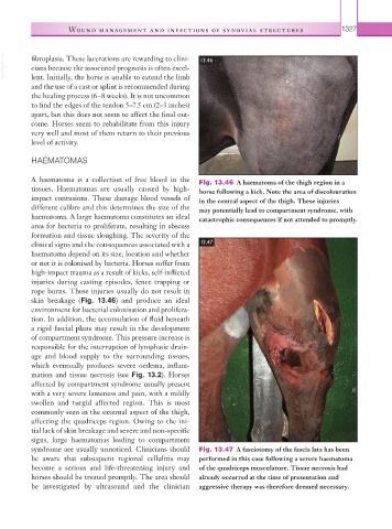
A haematoma is a collection of free blood in the Fig. 13.46 A haematoma of the thigh region in a
tissues. Haematomas are usually caused by high- horse following a kick. Note the area of discolouration
impact contusions. These damage blood vessels of in the central aspect of the thigh. These injuries
different calibre and this determines the size of the may potentially lead to compartment syndrome, with
haematoma. A large haematoma constitutes an ideal catastrophic consequences if not attended to promptly.
area for bacteria to proliferate, resulting in abscess
formation and tissue sloughing. The severity of the
clinical signs and the consequences associated with a 13.47
haematoma depend on its size, location and whether
or not it is colonised by bacteria. Horses suffer from
high-impact trauma as a result of kicks, self-inflicted
injuries during casting episodes, fence trapping or
rope burns. These injuries usually do not result in
skin breakage (Fig. 13.46) and produce an ideal
environment for bacterial colonisation and prolifera-
tion. In addition, the accumulation of fluid beneath
a rigid fascial plane may result in the development
of compartment syndrome. This pressure increase is
responsible for the interruption of lymphatic drain-
age and blood supply to the surrounding tissues,
which eventually produces severe oedema, inflam-
mation and tissue necrosis (see Fig. 13.2). Horses
affected by compartment syndrome usually present
with a very severe lameness and pain, with a mildly
swollen and turgid affected region. This is most
commonly seen in the external aspect of the thigh,
affecting the quadriceps region. Owing to the ini-
tial lack of skin breakage and severe and non-specific
signs, large haematomas leading to compartment
syndrome are usually unnoticed. Clinicians should Fig. 13.47 A fasciotomy of the fascia lata has been
be aware that subsequent regional cellulitis may performed in this case following a severe haematoma
become a serious and life-threatening injury and of the quadriceps musculature. Tissue necrosis had
horses should be treated promptly. The area should already occurred at the time of presentation and
be investigated by ultrasound and the clinician aggressive therapy was therefore deemed necessary.