
Page 1360 - Equine Clinical Medicine, Surgery and Reproduction, 2nd Edition
P. 1360
The foal 1335
VetBooks.ir 14.3 with antibody levels in the mare’s serum peaking at
9 days post partum. First foals, therefore, do not show
clinical signs. However, during subsequent pregnan-
cies, the secondary anamnestic response produces
significant antierythrocyte antibodies concentrated
in the colostrum, which puts the foal at risk. It is
also reported that tissue vaccines or transfusions may
sensitise a mare to genetically incompatible erythro-
cytes. Antibodies become concentrated in the colos-
trum during the last weeks of pregnancy and are
absorbed during the first 24 hours after birth. The
antibodies pass into the foal’s circulation and attach
themselves to the surface of the foal’s erythrocytes,
causing them to agglutinate and be removed by the
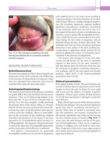
Fig. 14.3 The oral mucous membrane of a foal monocyte–phagocytic system, or to haemolyse intra-
showing petechiation due to an immune-mediated vascularly in the presence of complement.
thrombocytopenia. The disease only develops if: (1) the mare lacks
certain red cell factors; (2) the mare is repeatedly
exposed to that factor; (3) the mare conceives a
NEONATAL ISOERYTHROLYSIS foal that inherits that red cell factor from the sire;
(4) colostrum is produced containing antierythro-
Definition/overview cyte antibody; and (5) the foal ingests and absorbs
Neonatal isoerythorlysis (NI) is characterised by the antibody, which leads to the immune-mediated
destruction of the foal’s red blood cells (RBCs) due destruction of its red cells.
to the presence of alloantibodies of maternal origin
that were absorbed from colostrum. NI occurs in Clinical presentation
newborn foals within the first week of life. Foals appear healthy at birth. The speed of onset and
severity of clinical signs are dependent on the red cell
Aetiology/pathophysiology antigen involved (Aa and Qa being the most rapid
The disease occurs because of the presence of antibod- and severe) and the efficacy of transfer of passive
ies against RBCs that were absorbed with the mare’s immunity. Clinical signs usually develop between
colostrum. There are 32 blood-group antigens in the 12 and 48 hours post partum, although occasionally
horse, but 90% of cases of NI are associated with Aa up to 96 hours.
and Qa. Aa is the most antigenic, usually producing Early clinical signs may be subtle and non- specific.
the peracute form of the disease within 12–18 hours Foals become lethargic and yawn, become depressed,
of birth. Qa has not been associated with clinical dis- spend more time in recumbency and suck less fre-
ease but may cause a weak false positive when tests are quently. When they are excited or restrained, heart
carried out for the condition. There is a breed differ- rate and respiratory rate increase. Examination of
ence in the occurrence of the erythrocyte antigens the mucous membranes and sclera reveals varying,
that affects the prevalence of the disease. It has been but often marked, degrees of jaundice (Fig. 14.4). In
reported in 2% of Standardbreds and in less than 1% severe cases the urine becomes red due to the pres-
of Thoroughbreds. The frequency of isoimmunisation ence of haemoglobinuria. There can be a rapid pro-
is much lower than that of incompatible pregnancies, gression to collapse, coma and death in some cases.
and the mechanism of this difference is unknown. More frequently, in less severe cases, signs progress
The primary isoimmunisation by genetically more slowly, and in unusual cases the development of
incompatible erythrocytes occurs late in pregnancy, signs may be delayed.