
Page 619 - Equine Clinical Medicine, Surgery and Reproduction, 2nd Edition
P. 619
594 CHAPTER 3
VetBooks.ir • Caudally: a line drawn from the middle of the a small incision made. A 3–4 mm Steinmann pin in
orbit to the facial crest.
a chuck is used to drill through the bone into the
The boundaries of the frontal sinus are as follows: sinus. A catheter can then be introduced and a saline
lavage taken.
• Caudally: a line drawn from the Sinuscopy
temporomandibular joint to the midline. The entry points and preparation for sinuscopy
• Rostrally: the midpoint of a line drawn from (Fig. 3.5) are the same as for centesis except that a
the medial canthus of the eye to the nasoincisive larger pin size or trephine hole is required to provide
notch joined to the midline. access for the endoscope. A trephine hole into the
• Laterally: the medial canthus of the eye. frontal sinus also provides access to the caudal max-
illary sinus via the frontomaxillary opening and to
Percutaneous sinus centesis the ventral conchal sinus if forceps are used to create
This is a simple means of obtaining a lavage sam- an opening into this space.
ple from the sinuses in a standing, sedated horse.
The entry site for the caudal maxillary sinus is Tracheal and bronchoalveolar lavage
approximately 3–4 cm dorsal to the facial crest and Tracheal lavage (TL) and BAL are essential in all
approximately 3–4 cm rostral to the medial can- cases of LRT disease. TL and BAL samples are
thus. The entry site for the rostral maxillary sinus suitable for cytology, bacteriology/virology and
is approximately 3–4 cm rostral to this. The entry parasitology. As discharges pool in the trachea, TL
site for the frontal sinus is the midpoint of a line samples are representative of both lung fields, but
drawn from the medial canthus to the midline. generally they provide poor samples for cytology
Local anaesthetic is infiltrated subcutaneously and (cells are degenerate) and may not accurately reflect
current events in the lung. BAL samples provide an
accurate, current reflection of events in the lung,
3.5
but because only one segment of lung is sampled,
they are not representative of the whole lung. BAL
samples are thus suitable for generalised lung dis-
eases (e.g. equine asthma), but they may miss focal
abnormalities.
Tracheal lavage
TL is usually performed transendoscopically. There
is a small risk of bacterial contamination of transend-
oscopic samples from the nasal cavity and nasophar-
ynx, but this does not usually invalidate bacteriology
results because the quantity of contamination is
small. TL can be carried out percutaneously using
either a commercial kit or an intravenous cathe-
ter in cases where it is judged important to collect
the sample using a sterile collection method. The
endoscope is advanced to the mid-cervical trachea
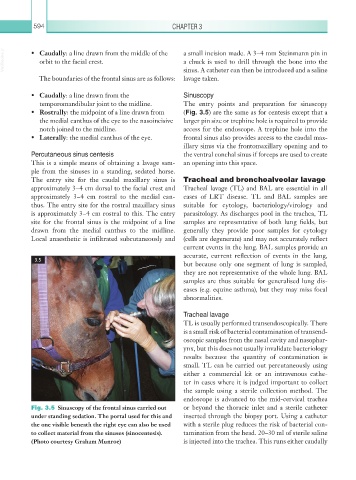
Fig. 3.5 Sinuscopy of the frontal sinus carried out or beyond the thoracic inlet and a sterile catheter
under standing sedation. The portal used for this and inserted through the biopsy port. Using a catheter
the one visible beneath the right eye can also be used with a sterile plug reduces the risk of bacterial con-
to collect material from the sinuses (sinocentesis). tamination from the head. 20–30 ml of sterile saline
(Photo courtesy Graham Munroe) is injected into the trachea. This runs either caudally