
Page 682 - Equine Clinical Medicine, Surgery and Reproduction, 2nd Edition
P. 682
Respir atory system: 3.2 Surgical conditions of the respir atory tr act 657
VetBooks.ir and rapidly developing subcutaneous emphysema Management
Any lacerations should be debrided and ventral
and oedema, and possible obstruction of the tra-
chea from external compression by swellings and/
or wall injury. drainage established. Closure of the wound is usu-
ally contraindicated and can result in severe absces-
sation with further compression and damage.
Differential diagnosis Healing by second intention should be encouraged
There is no specific differential diagnosis – more by regular careful wound management and antibi-
relevant are the potential involvement of other otic/anti-inflammatory medications. The trachea
structures. The common carotid artery lies dorso- usually heals well with second-intention healing.
lateral to the trachea, with the vagosympathetic In closed wounds pressure bandages, topical anti-
trunk deep to it. The oesophagus is usually left of, inflammatory measures and systemic antibiotic and
and dorsal to, the trachea. The recurrent laryn- anti-inflammatory medications are helpful. In severe
geal nerve lies dorsal to the trachea, in the deep respiratory obstruction a temporary tracheotomy
tissues of the neck. distal to the injury may be required. Primary sur-
gical repair of tracheal injuries is possible in some
Diagnosis cases. Suction decompression of the emphysema
Direct examination and sterile palpation of the using wide bore needles is also surprisingly effective
wound and endoscopic examination of the tra- in many cases.
chea will reveal the laceration from the outside
and inside (Fig. 3.103) and allow assessment of Prognosis
the damage. Radiography and ultrasonography The prognosis is fair. Generally, if treated appro-
will often reveal gas in the soft tissues of the neck priately and early, lacerations to trachea will heal
in such cases and possibly deformity of the tra- with no long-term problems. Extensive subcutane-
cheal outline or rings (Fig. 3.104). Subcutaneous ous emphysema can tract caudally into the thorax or
emphysema is common following lacerations to mediastinum leading to life-threatening complica-
the chest and pectoral area and does not necessar- tions. Tracheal lacerations can result in disturbance
ily indicate tracheal laceration. of the airflow and respiratory noise, due tracheal
stenosis, particularly if there is extensive damage to
the tracheal rings. However, this usually does not
result in significant airway stenosis.
3.103
3.104
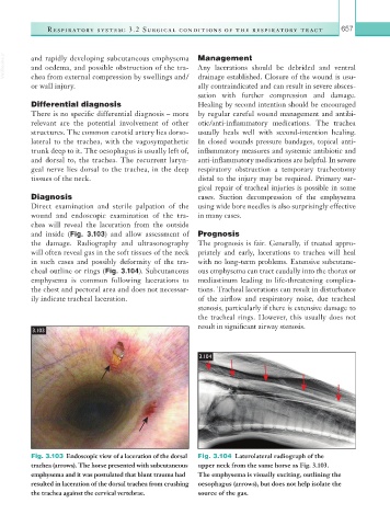
Fig. 3.103 Endoscopic view of a laceration of the dorsal Fig. 3.104 Laterolateral radiograph of the
trachea (arrows). The horse presented with subcutaneous upper neck from the same horse as Fig. 3.103.
emphysema and it was postulated that blunt trauma had The emphysema is visually exciting, outlining the
resulted in laceration of the dorsal trachea from crushing oesophagus (arrows), but does not help isolate the
the trachea against the cervical vertebrae. source of the gas.