
Page 980 - Equine Clinical Medicine, Surgery and Reproduction, 2nd Edition
P. 980
Urinary system 955
VetBooks.ir and sepsis, which most often include fever, weakness, 7.40
Foals may also show signs of concurrent infection
injected mucous membranes, diarrhoea and septic
arthritis. Severe electrolyte disturbances can cause
neurological abnormalities.
Differential diagnosis
Differential diagnoses include colic, pleuropneumo-
nia, sepsis, endotoxaemia, renal failure, neoplasia,
intestinal rupture and RTA.
Diagnosis
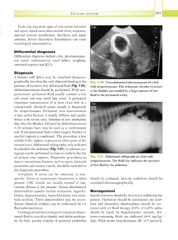
A bladder wall defect may be visualised ultrasono-
graphically but often the only abnormal finding is the Fig. 7.40 Transabdominal ultrasonogram of a foal
presence of excessive free abdominal fluid (Fig. 7.40). with uroperitoneum. The echogenic circular structure
Abdominocentesis should be performed. With uro- is the bladder surrounded by a large amount of free
peritoneum, peritoneal fluid usually contains a low fluid in the peritoneal cavity.
cell count and may smell like urine. A peritoneal
creatinine concentration of at least twice that of a 7.41
concurrently obtained serum sample is diagnostic
for uroperitoneum. Peritoneal urea determination
is less useful because it readily diffuses and equili-
brates with serum urea. Infusion of new methylene
blue into the bladder, followed by abdominocentesis
5–10 minutes later, may be used as a confirmatory
test. If the peritoneal fluid is blue-tinged, bladder or
urachal rupture is confirmed. The procedure is less
reliable if the rupture is present in other parts of the
urinary tract. Abdominal radiography only indicates
free fluid in the abdomen (Fig. 7.41). A contrast cys-
togram can be performed in foals to confirm the site
of urinary tract rupture. Diagnostic procedures to Fig. 7.41 Abdominal radiograph of a foal with
detect concomitant diseases, such as sepsis, bacterial uroperitoneum. The fluid line indicates the presence
peritonitis and urinary calculi, should be included in of free fluid in the abdomen.
the diagnostic procedure.
Urinalysis, if urine can be obtained, is non-
specific. Gross or microscopic haematuria is often should be evaluated, and the umbilicus should be
present. CBC results are usually normal if con- examined ultrasonographically.
current disease is not present. Serum biochemical
abnormalities usually include azotaemia, hyperka- Management
laemia, hyponatraemia, hypochloraemia and meta- Initial treatment should be directed at stabilising the
bolic acidosis. These abnormalities may be severe. patient. Hydration should be maintained and acid–
Serum chemical analysis can be influenced by i/v base and electrolyte abnormalities should be cor-
fluid administration. rected with i/v fluid therapy (0.9% or 0.45% saline
Cytological and bacteriological evaluation of peri- should be used). In hyperkalaemic animals, dex-
toneal fluid is crucial to identify and define peritoni- trose-containing fluids are indicated (4–8 mg/kg/
tis. In foals, passive transfer of maternal antibodies day). With severe hyperkalaemia (K >5.5 mmol/l),
+