
Page 1146 - Adams and Stashak's Lameness in Horses, 7th Edition
P. 1146
1112 Chapter 11
FARRIERY FOR COMMON HOOF PROBLEMS
VetBooks.ir Stephen e. O’Grady
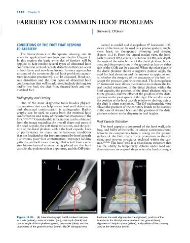
CONDITIONS OF THE FOOT THAT RESPOND Lateral to medial and dorsopalmar 0° horizontal (DP)
TO FARRIERY views of the foot can be used as a precise guide to imple
ment basic or therapeutic trimming and shoeing
The biomechanics of therapeutic shoeing and its (Figure 11.31). From the lateral medial view, the hoof‐
scientific application have been described in Chapter 8. pastern axis (HPA), center of rotation (COR), sole depth,
In this section the basic principles of farriery will be the angle of the solar border of the distal phalanx, break‐
applied to help resolve several types of abnormal hoof over, and the proportions of the ground surface on either
conformation or hoof capsule distortions that can occur side of the COR can be assessed. When the solar plane of
in both lame and non‐lame horses. Farriery approaches the distal phalanx shows a negative palmar angle, the
to some of the common clinical hoof problems encoun need for heel elevation and the amount to apply, as well
tered in equine practice will also be discussed. Hoof cap as whether the integrity of the structures of the heel will
sule distortion and the four types of abnormal hoof accept the pressure, can be determined. The dorsopalmar
conformation that will be addressed include the long toe 0° horizontal view allows the clinician to evaluate the lat
and/or low heel, the club foot, sheared heels and mis eral medial orientation of the distal phalanx within the
matched feet. hoof capsule, the position of the distal phalanx relative
to the ground, and the effect of the position of the distal
Radiography and Farriery phalanx on the joint spaces of the digit. The need to assess
the position of the hoof capsule relative to the long axis of
One of the main diagnostic tools besides physical the digit is often overlooked. The DP radiographic view
examination that can help assess hoof wall distortions allows the position of the coronary bands to be assessed
and abnormal conformation is radiography. Radio in the case of sheared heels and the position of the distal
graphy can be used to assess both the external hoof phalanx relative to the disparity in heel heights.
conformation and many of the internal structures of the
foot. 4,5,22,31,39 Considerable information can be obtained
from the image regarding the overall shape and mass of Hoof Capsule Distortion
the hoof capsule, the soft tissue structures, and the posi The hoof capsule is composed of the hoof wall, sole,
tion of the distal phalanx within the hoof capsule. Lack frog, and bulbs of the heels. Its unique continuous bond
of performance or many subtle lameness conditions between its components form a casing on the ground
that are localized to the foot are caused by hoof capsule surface of the limb that affords protection to the soft
distortions, poor foot conformation, improper landing tissue and osseous structures enclosed within the cap
patterns, and soft tissue damage that result from exces sule. 13,33,36 The hoof wall is a viscoelastic structure that
sive biomechanical stresses being placed on the hoof has the ability to temporarily deform under load and
capsule, the podotrochlear apparatus, and the DIP joint. then return to its original shape when the load or weight
A B
Figure 11.31. (A) Lateral radiograph that illustrates hoof‐pas- illustrates the axial alignment of the digit (red), position of the
tern axis (yellow), center of rotation (red), sole depth (black) and foramina in the distal phalanx relative to the ground (blue),
palmar angle of distal phalanx (green), break‐over (blue), and the alignment of the joint space (yellow), and position of the coronary
proportions of the ground surface (white). (B) DP radiograph that band at the heel bulbs (white).