
Page 1182 - Adams and Stashak's Lameness in Horses, 7th Edition
P. 1182
1148 Chapter 12
it is difficult to secure the splints to the limb if the
bandage on the distal aspect of the metatarsus is too
VetBooks.ir of padding each compressed with brown gauze or elastic
bulky. Robert‐Jones bandages consist of multiple layers
tape, and each layer should be applied more tightly than
the previous one. 14
Tarsus and Tibia
Fractures in the tarsus and tibia are particularly dif
ficult to adequately stabilize because of the reciprocal
apparatus and its effect on joint motion in the tarsus
and stifle. Fractures in this location tend to collapse
3,4
when the stifle flexes, and the tarsus remains in a fixed
position. The main principle for stabilization is similar
to that of the radius, which primarily involves preventing
abduction of the distal aspect of the limb (Figure 12.11).
There is very little muscle coverage of the medial aspect
of the tibia, and this is the site that is prone to becoming
open. To prevent abduction, a single laterally placed
splint that is bent to follow the angulation of the limb
and extends proximally above the stifle joint works
well. The splint is most effective if it is bent back on
itself to mirror the hindlimb contour back to the ground,
creating a double splint. The splint is applied over a full‐
limb Robert‐Jones bandage and is best made of light
weight metal such as aluminum (Figure 12.12) that can
be bent into the proper position, similar to the lateral
support of a Schroeder‐Thomas splint. An alternative to
the metal splint is a wide board (15–20 cm) that extends
from the ground to the ilium and is applied to the lateral
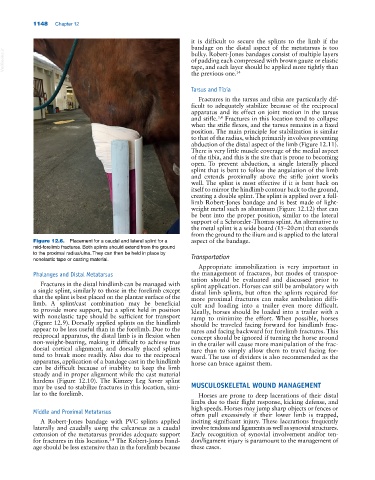
Figure 12.6. Placement for a caudal and lateral splint for a aspect of the bandage.
mid‐forelimb fractures. Both splints should extend from the ground
to the proximal radius/ulna. They can then be held in place by Transportation
nonelastic tape or casting material.
Appropriate immobilization is very important in
Phalanges and Distal Metatarsus the management of fractures, but modes of transpor
tation should be evaluated and discussed prior to
Fractures in the distal hindlimb can be managed with splint application. Horses can still be ambulatory with
a single splint, similarly to those in the forelimb except distal limb splints, but often the splints required for
that the splint is best placed on the plantar surface of the more proximal fractures can make ambulation diffi
limb. A splint/cast combination may be beneficial cult and loading into a trailer even more difficult.
to provide more support, but a splint held in position Ideally, horses should be loaded into a trailer with a
with nonelastic tape should be sufficient for transport ramp to minimize the effort. When possible, horses
(Figure 12.9). Dorsally applied splints on the hindlimb should be traveled facing forward for hindlimb frac
appear to be less useful than in the forelimb. Due to the tures and facing backward for forelimb fractures. This
reciprocal apparatus, the distal limb is in flexion when concept should be ignored if turning the horse around
non‐weight‐bearing, making it difficult to achieve true in the trailer will cause more manipulation of the frac
dorsal cortical alignment, and dorsally placed splints ture than to simply allow them to travel facing for
tend to break more readily. Also due to the reciprocal ward. The use of dividers is also recommended as the
apparatus, application of a bandage cast in the hindlimb horse can brace against them.
can be difficult because of inability to keep the limb
steady and in proper alignment while the cast material
hardens (Figure 12.10). The Kimzey Leg Saver splint
may be used to stabilize fractures in this location, simi MUSCULOSKELETAL WOUND MANAGEMENT
lar to the forelimb. Horses are prone to deep lacerations of their distal
limbs due to their flight response, kicking defense, and
high speeds. Horses may jump sharp objects or fences or
Middle and Proximal Metatarsus
often pull excessively if their lower limb is trapped,
A Robert‐Jones bandage with PVC splints applied inciting significant injury. These lacerations frequently
laterally and caudally using the calcaneus as a caudal involve tendons and ligaments as well as synovial structures.
extension of the metatarsus provides adequate support Early recognition of synovial involvement and/or ten
for fractures in this location. The Robert‐Jones band don/ligament injury is paramount to the management of
3,4
age should be less extensive than in the forelimb because these cases.