
Page 296 - Adams and Stashak's Lameness in Horses, 7th Edition
P. 296
VetBooks.ir
a
15
14 1
13
12 2
6
11 3
9
10
7 4
9
8
7
6
b
5
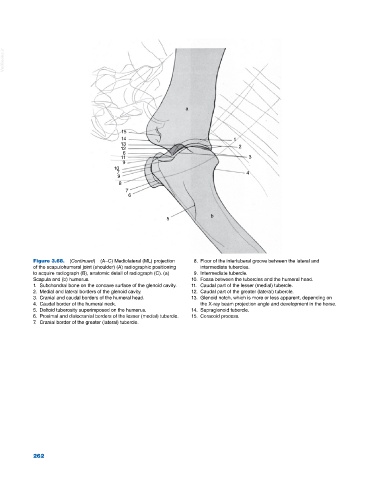
Figure 3.68. (Continued) (A–C) Mediolateral (ML) projection 8. Floor of the intertuberal groove between the lateral and
of the scapulohumeral joint (shoulder) (A) radiographic positioning intermediate tubercles.
to acquire radiograph (B), anatomic detail of radiograph (C). (a) 9. Intermediate tubercle.
Scapula and (b) humerus. 10. Fossa between the tubercles and the humeral head.
1. Subchondral bone on the concave surface of the glenoid cavity. 11. Caudal part of the lesser (medial) tubercle.
2. Medial and lateral borders of the glenoid cavity. 12. Caudal part of the greater (lateral) tubercle.
3. Cranial and caudal borders of the humeral head. 13. Glenoid notch, which is more or less apparent, depending on
4. Caudal border of the humeral neck. the X‐ray beam projection angle and development in the horse.
5. Deltoid tuberosity superimposed on the humerus. 14. Supraglenoid tubercle.
6. Proximal and distocranial borders of the lesser (medial) tubercle. 15. Coracoid process.
7. Cranial border of the greater (lateral) tubercle.
262