
Page 298 - Adams and Stashak's Lameness in Horses, 7th Edition
P. 298
13 1
VetBooks.ir 11 2
3
a 4
5
b c
12
6
11
4
10
5
9
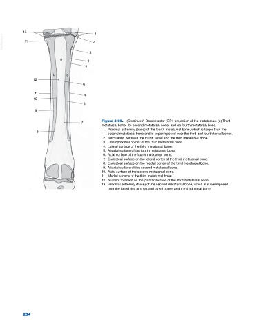
Figure 3.69. (Continued) Dorsoplantar (DPl) projection of the metatarsus. (a) Third
7
metatarsal bone, (b) second metatarsal bone, and (c) fourth metatarsal bone.
1. Proximal extremity (base) of the fourth metatarsal bone, which is larger than the
8
second metatarsal bone and is superimposed over the third and fourth tarsal bones.
2. Articulation between the fourth tarsal and the third metatarsal bone.
3. Lateroproximal border of the third metatarsal bone.
4. Lateral surface of the third metatarsal bone.
5. Abaxial surface of the fourth metatarsal bone.
6. Axial surface of the fourth metatarsal bone.
7. Endosteal surface on the lateral cortex of the third metatarsal bone.
8. Endosteal surface on the medial cortex of the third metatarsal bone.
9. Abaxial surface of the second metatarsal bone.
10. Axial surface of the second metatarsal bone.
11. Medial surface of the third metatarsal bone.
12. Nutrient foramen on the plantar surface of the third metatarsal bone.
13. Proximal extremity (base) of the second metatarsal bone, which is superimposed
over the fused first and second tarsal bones and the third tarsal bone.
264