
Page 340 - Adams and Stashak's Lameness in Horses, 7th Edition
P. 340
VetBooks.ir
B
A
C D
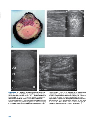
Figure 3.91. In Z1B the palmar metacarpal structures appear much footprint (the SDF and DDF are not as wide as the SL) and the creation
as they did in zone 1A. The SDFT appears to flatten palmar to dorsal, of edge artifacts and acoustic enhancement artifacts from more
and the DDFT becomes more rounded. The ICL becomes more inclined superficial structures (tendons and vessels) that can make evaluation of
toward the DDFT. Fibers of the SL become more pronounced, and this the SL difficult (C). Angled contrast ultrasound (ACUST) utilizes off‐
structure widens medial to lateral (A). The SDF and DDF tendons angled and non‐weight‐bearing imaging of the proximal suspensory to
should be evaluated with the focal zones placed at the appropriate level allow visualization of the muscle and fat bundles within the origin of the
and often with a standoff pad in place (B). However, ultrasound imaging SL not normally evident in standard transverse ultrasound imaging of
of the suspensory ligament in this area is often difficult due to a limited this area (D). Source: US images courtesy of Dr. Caitlyn Horne.
306