
Page 353 - Adams and Stashak's Lameness in Horses, 7th Edition
P. 353
VetBooks.ir
B
A
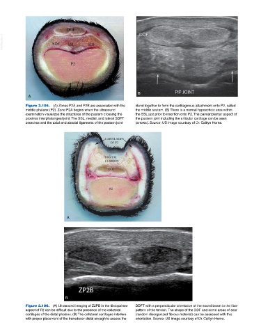
Figure 3.105. (A) Zones P2A and P2B are associated with the blend together to form the cartilaginous attachment onto P2, called
middle phalanx (P2). Zone P2A begins when the ultrasound the middle scutum. (B) There is a normal hypoechoic area within
examination visualizes the structures of the pastern crossing the the SSL just prior to insertion onto P2. The palmar/plantar aspect of
proximal interphalangeal joint. The SSL, medial, and lateral SDFT the pastern joint including the articular cartilage can be seen
branches and the axial and abaxial ligaments of the pastern joint (arrows). Source: US image courtesy of Dr. Caitlyn Horne.
A
B
Figure 3.106. (A) Ultrasound imaging of Z2PB in the distopalmar DDFT with a perpendicular orientation of the sound beam to the fiber
aspect of P2 can be difficult due to the presence of the collateral pattern of the tendon. The shape of the DDF and some areas of scar
cartilages of the distal phalanx. (B) The collateral cartilages interfere (random disorganized fibrous material) can be assessed with this
with proper placement of the transducer distal enough to assess the orientation. Source: US image courtesy of Dr. Caitlyn Horne.