
Page 377 - Adams and Stashak's Lameness in Horses, 7th Edition
P. 377
Diagnostic Imaging 343
administered intravenously and images made as soon as the personnel even further (ALARA). Radiation safety
radiopharmaceutical equilibrium in the extracellular rules may vary from state to state; therefore, the practi
VetBooks.ir scanning agents is recommended. 99m Tc‐RBC is another and comply with the specific state regulations.
tioner should consult the local radiation safety officer
space is achieved. A similar dose to that of the bone‐
alternative for evaluating blood perfusion to soft tissues
without the risk of bone uptake overlap. 83
Phase 3, known as the delayed or bone phase, occurs IMAGING EQUIPMENT
several hours later when approximately 50% of the
injected radiotracer has attached to the bone. The The gamma camera contains a collimator made of
remainder of the tracer is excreted by the kidneys in the small holes in a lead plate that allows only perpendicu
first 1 or 2 urine voids’ postinjection. The uptake pat lar γ‐rays through. This reduces scatter, thereby improving
tern of normal bone is quite predictable and is described image resolution. The γ‐rays interact with a fluorescent
later in this chapter. The diaphysis of long bones has the crystal (a thallium‐activated sodium iodide crystal is
least uptake, and greatest uptake of the tracer occurs in commonly used), changing the γ‐energy to light pho
the juxtaphyseal and subchondral bone in normal sub tons. The light photons interact with a photocathode,
jects. Increased radiotracer by or near the joints during generating electrons that are amplified by an array of
the delayed (bone) phase has been related to osteoarthri photomultiplier tubes. The x–y coordinates of the elec
tis (OA), various enthesopathies, periarticular bone scle trons are then recorded, and the image is reconstructed.
rosis, septic arthritis, etc. These changes from the normal The resultant image represents the geographic distribu
radiotracer pattern are discussed later in this chapter. tion of the radiotracer in the horse. Images can be
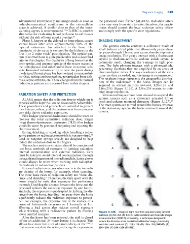
acquired in several matrices (64 × 64, 128 × 128 and
256 × 256) (Figure 3.126). A 256 × 256 matrix to opti
mize image resolution.
RADIATION SAFETY AND PROTECTION Various techniques have been devised to suspend the
ALARA means that the radiation dose to which one is gamma camera such as a stationary actuated lift or
69
exposed will be kept “As Low As Reasonably Achievable.” track‐and‐column mounted detectors (Figure 3.127).
These procedures and protocols are intended to protect The tract system can moved around the horses, whereas
the clinician, others, and the environment from unneces in the stationary system, the horse is moved around the
sary risks due to radiation exposures. camera.
Film badges (personal dosimeters) should be worn to
monitor the total cumulative radiation dose. Finger
(ring) thermoluminescent dosimetry (TLD) film badges
should be worn by personnel preparing and injecting the
pharmaceutical.
Eating, drinking, or smoking while handling a radio
active patient or radioactive materials is not permitted.
98
Lead or tungsten syringe shields are designed to help
reduce the radiation dose to the fingers.
The nuclear medicine clinician should be conscious of
two basic methods of exposure to ionizing radiation: A B
internal contamination and external radiation. Care
must be taken to avoid internal contamination through
the accidental ingestion of the radionuclide. Latex gloves
should always be worn when working with radiophar
maceuticals or radioactive patients.
External radiation occurs when one is in the immedi
ate vicinity of the horse, for example, when scanning.
The three basic rules of radiation safety are “time, dis C D
tance, and shielding.” Therefore, the time spent with the
horse should be only that necessary for performing
the study. Doubling the distance between the horse and the
personnel reduces the radiation exposure by one fourth.
Similarly, the exposure is quadrupled by halving the dis
tance from the horse. Standing 1 m away from the horse
significantly reduces the radiation exposure to person
nel. For example, the exposure rate at the surface of a
horse of 6.6 mrem/h decreases to 1.3 mrem/h at 1 m. E F
Wearing a lead apron also reduces overall exposure
while working with a radioactive patient by filtering Figure 3.126. Image of right front foot acquired using several
lower emitted energies. matrices. (A) 64 × 64. (B) 64 × 64 with statistical and heuristic image
After the horse has been released, the stall is closed noise extraction (SHINE) processing, a technique designed to
off for an additional 24 hours before it is cleaned. This reduce the Poisson noise contained within the image, preserving
allows four more half‐lives of natural decay of the 99m Tc resolution and contrast. (C) 128 × 128. (D) 128 × 128 (SHINE). (E)
that was excreted via the urine, reducing the exposure to 256 × 256. (F) 256 × 256 (SHINE).