
Page 526 - Adams and Stashak's Lameness in Horses, 7th Edition
P. 526
492 Chapter 4
loss of rigidity due to cytoskeletal dysregulation/col- PATHOGENESIS: STRUCTURAL CONSIDERATIONS
lapse secondary to aberrant epithelial cell signaling. OF THE EQUINE DIGIT
VetBooks.ir 48 hours in the EHC model (likely due to the supra- borne by the two anatomical structures to which the
Whereas these lamellar changes take place over
The stresses on the lamellae are related to the stresses
physiologic blood insulin concentrations maintained),
43
the changes can occur gradually over months to years lamellae attach, the hoof wall and the distal phalanx.
in the clinical patient. Different stresses occur due to both weight‐bearing and
In SLL, a recently introduced model in which a shoe locomotion. The three primary forces affecting the
with a V‐shaped insert on one limb causes excessive lamellae include:
weight‐bearing on the other limb has started to shed 1. The downward force of the horse’s weight through
light on the events preceding lamellar failure. This the distal phalanx
establishment of a model for SLL was critical due to 2. The torque (or moment) around the distal inter-
the fact that clinical cases of SLL are characterized by phalangeal (DIP) joint created by the ground reac-
a rapid onset of severe lamellar injury/separation, mak- tion force (the force exerted by the ground on the
ing it difficult to assess early changes in the disease digit; also termed extensor moment)
process. Whereas the first study using this model sug- 3. The tension of the deep digital flexor tendon (DDFT)
gested an increase in lamellar hypoxia in SLL due to exerted on the caudal aspect of the distal phalanx
increased concentrations of a protein induced by (also termed the flexor moment; Figure 4.65)
hypoxia, a more recent study using the same SLL
33
model in which microdialysis probes were placed in the It has been demonstrated in a horse with healthy feet
lamellae more clearly indicated a decrease in lamellar that the sole of the foot can support the weight of the
blood flow starting approximately 48 hours after the limb (i.e. counteracting the downward force on the dis-
onset of preferential weight‐bearing (A. van Eps, tal phalanx from the horse’s weight), but it is unknown
unpublished data). On histology of lamellar samples how the forces associated with weight‐bearing are dis-
harvested at 96 hours of preferential weight‐bearing, tributed from the sole to the distal phalanx. More spe-
both lamellar stretching and dysadhesion were present cifically, the question is how much of the weight borne
(Belknap and van Eps, unpublished data). As the by the sole is directly transferred to the distal phalanx
decreased blood flow is likely due to physical factors and how much is redirected through the lamellae to the
such as a lack of movement, it is unlikely that phar- distal phalanx. The following discussion assumes that
23
macologic vasodilation will be effective in preventing there is some direct support of the distal phalanx by the
or treating SLL. The breakdown in lamellar epithelial sole, but this remains unproven. In the horse shod with
structure in SLL may not only be due to the direct standard shoeing that is standing on a firm flat surface,
effects of hypoxia on cell metabolism; we have also all the stress of weight‐bearing is borne by the lamellae
recently established that the same growth factor‐related because the only relationship between the foot and the
signaling as discussed above is occurring in the lamel- ground surface is through the part of the foot contacting
lar epithelial cells in the SLL model; this is most likely the shoe, the distal hoof wall. On the assumption that
stimulated by factors activated due to hypoxia (Belknap significant weight is transferred directly from the sole to
and van Eps, unpublished data). Thus, in all three types the distal phalanx, then a decrease in solar support of the
of laminitis, the induction of this same growth factor‐ distal phalanx will be exacerbated by the elevation of
related signaling may lead to lamellar failure through the sole off the ground surface due to the thickness of
disruption of epithelial cell cytoskeletal dynamics and the shoe; in the unshod foot, the character of the ground
adhesion to the underlying matrix. surface would therefore affect the amount of stress
G
g G g
d D
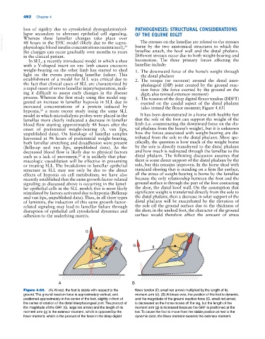
A B
Figure 4.65. (A) At rest, the foot is stable with respect to the flexor tendon (D, small red arrow) multiplied by the length of its
ground. The ground reaction force is approximately vertical, and moment arm (d). (B) At break‐over, the position of the foot is dynamic,
positioned approximately in the center of the foot, slightly in front of and the magnitude of the ground reaction force (G, small red arrow)
the center of rotation of the distal interphalangeal joint. The product of is decreased as the horse moves off the leg, but the length of the
the magnitude of the GRF (G, large red arrow) and the length of its moment arm (g) is increased because the GRF is positioned at the
moment arm (g) is the extensor moment, which is opposed by the toe. To cause the foot to move from the stable position at rest to the
flexor moment, which is the product of the force in the deep digital dynamic state, the flexor moment exceeds the extensor moment.