
Page 527 - Adams and Stashak's Lameness in Horses, 7th Edition
P. 527
Lameness of the Distal Limb 493
borne by the lamellae. On a hard surface (including arti-
ficial surfaces commonly used in stables), only the distal
VetBooks.ir results in a similar great degree of stress on the lamellae
hoof wall and adjacent sole contact the ground. This
similar to the foot with standard shoes. This is due to the
combined effects of concentration of stress through
the hoof wall to the lamellae and the lack of support of
the distal surface of the distal phalanx. In both of these
instances, the distal phalanx can be thought of as being
suspended within the foot by the lamellar attachments
to the hoof wall. When placed on a softer surface such
as sand (or possibly an elastic flooring material), the
entire solar surface can be engaged to directly support
the distal phalanx in addition to being suspended by the A
lamellar attachments to the hoof wall. This helps to dis-
tribute the digital support through both anatomic struc-
tures and thus decrease the stress on the lamellae. As is
37
discussed later, addition of putty to the solar surface in
the shod horse has the same end result.
At rest, the center of weight‐bearing is approxi-
mately at the center of the ground surface of the foot.
During the majority of locomotion, the stresses associ-
ated with weight‐bearing are also centered on the
ground surface of the foot. However, during the break‐
over stage of the stride, ground reaction forces are
localized to the toe of the foot, resulting in an increase
in the length of the moment arm (and hence the torque
or moment; see Figure 4.65) and thus the stress on the
dorsal lamellae.
In a healthy horse, the strength of the lamellae greatly
exceeds the stresses applied, thereby allowing the distal
phalanx to be retained in its normal position. However, B
in horses with laminitis, the lamellar attachments to the
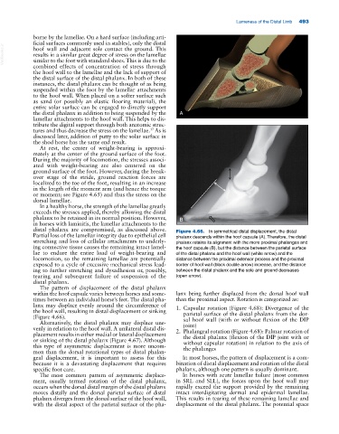
distal phalanx are compromised, as discussed above. Figure 4.66. In symmetrical distal displacement, the distal
Partial loss of the lamellar integrity due to epithelial cell phalanx descends within the hoof capsule (A). Therefore, the distal
stretching and loss of cellular attachments to underly- phalanx retains its alignment with the more proximal phalanges and
ing connective tissue causes the remaining intact lamel- the hoof capsule (B), but the distance between the parietal surface
lae to endure the entire load of weight‐bearing and of the distal phalanx and the hoof wall (white arrow) and the
locomotion, so the remaining lamellae are potentially distance between the proximal extensor process and the proximal
exposed to a cycle of excessive mechanical stress lead- border of hoof wall (black double arrow) increase, and the distance
ing to further stretching and dysadhesion or, possibly, between the distal phalanx and the sole and ground decreases
tearing and subsequent failure of suspension of the (open arrow).
distal phalanx.
The pattern of displacement of the distal phalanx
within the hoof capsule varies between horses and some- lanx being further displaced from the dorsal hoof wall
times between an individual horse’s feet. The distal pha- than the proximal aspect. Rotation is categorized as:
lanx may displace evenly around the circumference of 1. Capsular rotation (Figure 4.68): Divergence of the
the hoof wall, resulting in distal displacement or sinking parietal surface of the distal phalanx from the dor-
(Figure 4.66). sal hoof wall (with or without flexion of the DIP
Alternatively, the distal phalanx may displace une-
joint)
venly in relation to the hoof wall. A unilateral distal dis- 2. Phalangeal rotation (Figure 4.68): Palmar rotation of
placement results in either medial or lateral displacement the distal phalanx (flexion of the DIP joint with or
or sinking of the distal phalanx (Figure 4.67). Although without capsular rotation) in relation to the axis of
this type of asymmetric displacement is more uncom- the phalanges
mon than the dorsal rotational types of distal phalan-
geal displacement, it is important to assess for this In most horses, the pattern of displacement is a com-
because it is a devastating displacement that requires bination of distal displacement and rotation of the distal
specific foot care. phalanx, although one pattern is usually dominant.
The most common pattern of asymmetric displace- In horses with acute lamellar failure (most common
ment, usually termed rotation of the distal phalanx, in SRL and SLL), the forces upon the hoof wall may
occurs when the dorsal distal margin of the distal phalanx rapidly exceed the support provided by the remaining
moves distally and the dorsal parietal surface of distal intact interdigitating dermal and epidermal lamellae.
phalanx diverges from the dorsal surface of the hoof wall, This results in tearing of these remaining lamellae and
with the distal aspect of the parietal surface of the pha- displacement of the distal phalanx. The potential space