
Page 542 - Adams and Stashak's Lameness in Horses, 7th Edition
P. 542
508 Chapter 4
(Figure 4.82D). Based on these responses, the surgery is Sepsis of the distal phalanx can be difficult to both
primarily indicated in horses with: diagnose and treat. It is difficult to diagnose because the
VetBooks.ir 1. Early chronic laminitis that continues to rotate with sepsis closely resemble those associated with pro-
radiographic changes in the distal phalanx associated
despite all other measures taken
longed inflammation, and therefore, septic and nonseptic
2. Intractable pain originating from the dorsal sole and
wall despite stabilization and shoeing inflammation is very hard to distinguish. In the majority
of subsolar sepsis cases, the sepsis does not involve the
3. Secondary flexural deformities
bone. However, direct contact of bone after inserting a
It does not appear to consistently benefit horses with probe in a draining track conclusively identifies exposure
distal displacement and rarely if ever benefits the horse of the bone to sepsis and is highly suggestive of septic
with unilateral distal displacement of the distal phalanx. osteitis (combined with radiographs indicative of severe
The surgery may be performed in the mid‐metacarpal focal lysis at point of probe contact).
region (Figure 4.82A) or in the midpastern region. 1,69 The In the authors’ experience, septic osteitis of the distal
surgery is easier to perform in the mid‐metacarpal region. phalanx is much more refractory to treatment in horses
Additionally, should a second tenotomy be necessary, it is with laminitis than in horses in which the sepsis occurred
preferable to perform the proximal one first because for another reason. Additionally, surgery, usually con-
adhesions may limit the effectiveness of the second sur- sisting of curettage of the suspect bone, exposes the sur-
gery in the metacarpal region if the first surgery was per- face of the distal phalanx in a horse that did not have
formed in the pastern. Tenotomy in the midpastern septic osteitis and increases the likelihood that the horse
appears to provide greater mobility of the foot about the will develop septic osteitis. Therefore, infection should
DIP joint, but may also cause more instability of the joint. be treated as superficial unless drainage can be directly
The tendon may be cut immediately before or after linked to the bone or prolonged treatment fails to resolve
corrective trimming and shoeing is performed (it is prob- it and septic osteitis is the best explanation for the con-
ably best to perform the surgery first to address possible
DIP subluxation following DDFT). Because tissue repair
at the tenotomy site occurs fairly rapidly and will inhibit
any further realignment, it is imperative to obtain the
best realignment of the distal phalanx with the ground
and phalanges as soon as possible after surgery (i.e.
within hours to days). To counter the disadvantages of
the surgery, it is advisable to perform radiographs while
shoeing immediately following tenotomy of the DDFT to
assess both the adequacy of the realignment of the distal
phalanx to the ground surface and, importantly, the
degree of subluxation of the DIP joint (indicated by both
dorsal displacement of the extensor process of the distal
phalanx away from the middle phalanx [arrow,
Figure 4.82D] and by palmar displacement of distal
articular surface of the middle phalanx in relationship to
the articular surface of the distal phalanx [line,
Figure 4.82D]). Application of increasing degrees of heel
elevation can be assessed radiographically until the sub-
luxation is resolved. If radiographic assessment is not
available post‐tenotomy, the horse should be shod with
mild heel extension and elevation (approximately 3°).
Drainage and Debridement
Digital sepsis is a well‐recognized complication asso-
ciated with laminitis. Drainage may occur at the coro-
nary band or through the dorsal sole. In most horses, the
infection is confined to the soft tissues of the hoof, but,
occasionally, the infection may involve the distal pha-
lanx. If the hoof capsule of the sole is removed, the solar
dermis usually prolapses, and the prolapsed tissue is
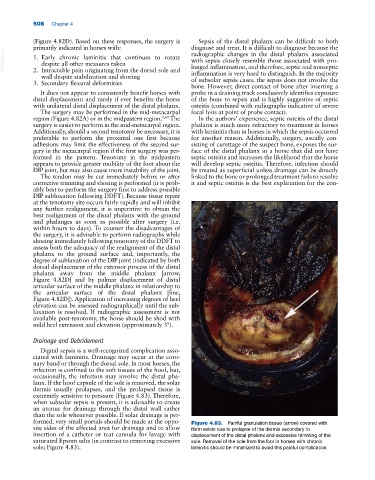
extremely sensitive to pressure (Figure 4.83). Therefore,
when subsolar sepsis is present, it is advisable to create
an avenue for drainage through the distal wall rather
than the sole whenever possible. If solar drainage is per-
formed, very small portals should be made at the oppo- Figure 4.83. Painful granulation tissue (arrow) covered with
site sides of the affected area for drainage and to allow fibrin exists due to prolapse of the dermis secondary to
insertion of a catheter or teat cannula for lavage with displacement of the distal phalanx and excessive trimming of the
saturated Epsom salts (in contrast to removing excessive sole. Removal of the sole from the foot in horses with chronic
sole; Figure 4.83). laminitis should be minimized to avoid this painful complication.