
Page 614 - Adams and Stashak's Lameness in Horses, 7th Edition
P. 614
580 Chapter 4
ticularly if the horse is used for performances other than The percentage of muscle in each region has been
17
racing. The prognosis for open comminuted splint bone identified; Standardbreds had 40% more muscle in their
VetBooks.ir mately one‐third of the proximal splint remains. The this may be associated with biomechanical differences in
SL than Thoroughbreds, and it has been suggested that
fractures is good to excellent with surgery if approxi-
gait between the two breeds or genetic factors. In vitro
prognosis for open comminuted fractures of the proximal
splint is more guarded, and about 60% return to perfor- strength testing of the suspensory apparatus in training
mance without lameness. These cases can be complicated and resting horses suggests that there is an increase of
by sequestration, osteomyelitis, and infectious arthritis. strength with training. The absolute load to failure in a
single load‐to‐failure compression test was higher in
horses that had been in racehorse training, and failure in
ENOSTOSIS‐LIKE LESIONS the trained group was usually by fracture of a proximal
sesamoid bone. In the untrained group, the SL failed. 22
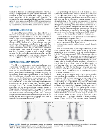
Enostosis‐like lesions (ELLs) have been identified in Injuries to the SL can be divided into three areas:
the third metacarpal and metatarsal bones on nuclear
1,9
scintigraphic examinations. Lesions are identified as 1. Lesions restricted to the proximal one‐third (proxi-
areas of focal, moderate to intense increased radiophar- mal suspensory desmitis [PSD])
maceutical uptake (IRU) in the medullary cavity 2. Lesions in the middle one‐third, sometimes extending
(Figure 4.158). Radiography of the affected bone may into the proximal third (body lesions)
reveal areas of increased radiodensity in the medullary 3. Lesions in the medial and/or lateral branch (branch
cavity, often near the nutrient foramen. ELLs in the met- lesions)
acarpal/metatarsal bones may or may not be associated PSD, or inflammation of the origin of the SL, is the
with lameness. Other causes of lameness should be ruled most common cause of soft tissue injury to the limbs,
out first. Treatment consists of stall or small paddock comprising approximately 30% of tendon/ligament
turnout for 3 months. Prognosis is generally good for injuries and approximately 60% of soft tissue injuries
143
return to athleticism; however, horses with more than that localize to the proximal metacarpus/metatarsus
one ELL appear to have a decreased prognosis. 1
(MC/MT). PSD occurs most commonly in sport horses,
22
such as event horses, jumpers, dressage horses, and race-
SUSPENSORY LIGAMENT DESMITIS horses. The hindlimbs are more frequently affected than
forelimbs and have a lower success rate of returning to
The SL is predominantly a strong, tendinous band performance after rest and convalescence: 69% for
containing variable amounts of muscular tissue hindlimbs vs. 80% for forelimbs. Overall, lameness
22
(2–11%). 39,42 It originates from the palmar carpal liga- associated with the origin of the SL occurs in about 5%
ment and the proximal palmar surface of the third meta- of horses (36 of 1,094 horses and 31 of 638 horses) with
carpal bone in the forelimb and descends between the a forelimb lameness. 36,37,39,42
second and fourth metacarpal bones. In the hindlimb, Injury can be exclusively within the ligament, involve
the SL originates primarily from the proximoplantar tearing of the Sharpey fibers at the origin of the SL, or be
aspect of the third metatarsal bone. An accessory liga- associated with avulsion fracture(s) of the origin of the
ment originating from the plantar fourth tarsal bone SL that involves the proximal MC/MT. High PSD and
also joins the SL. In the distal metacarpus/metatarsus it acute tearing of Sharpey fibers may present with similar
divides into two branches that insert on the proximal signs as those described for blind splints and desmitis
sesamoid bones. The extensor branches pass obliquely of the carpal (distal) check ligament. In chronic cases of
dorsad to join the common digital extensor tendon in tearing of the origin of the SL, radiographic evidence
the proximal phalangeal region. The suspensory appara- of bone resorption and surrounding sclerosis is often
tus is continued distally as the straight, oblique, cruciate, present. With avulsion fracture of the origin of the SL,
and short distal sesamoidean ligaments. The SL phyloge- either the lateral or medial branch may be involved in
netically represents the median interosseous muscle and one or both limbs and will likely be noted on radio-
is also known as the interosseous muscle or proximal graphs or seen as a displaced piece of bone on ultra-
(superior) sesamoidean ligament. 36,37,39,42 sound (Figure 4.159). This lameness most frequently
Left Hind Lat Right Hind Lat PD Hinds Fetlocks
L R
ALL FOUR
ALL FOUR
Figure 4.158. Lateral and dorsopalmar nuclear scintigraphic uptake in the proximal medullary cavity of the right front limb (arrows)
images of the metacarpal regions in a horse with right front lameness. consistent with an enostosis‐like lesion (ELL).
There is a focal, intense area of increased radiopharmaceutical