
Page 619 - Adams and Stashak's Lameness in Horses, 7th Edition
P. 619
Lameness of the Distal Limb 585
SUPERFICIAL DIGITAL FLEXOR (SDF)
TENDINITIS (BOWED TENDON)
VetBooks.ir working at high speeds including Thoroughbred and
Tendinitis of the SDFT is a common injury in horses
Standardbred racehorses. Thoroughbred racehorses
have a reported incidence of 7%–43%. 34,116 Superficial
digital flexor (SDF) tendinitis can also occur in other
performance horses including show jumpers, event
horses, dressage horses, and Quarter Horses. Unlike SL
injury, SDFT injury is almost exclusively a forelimb
problem. Lesions can range from peritendinous inflam-
mation and pain without structural damage to complete
tendon rupture. Tendinitis usually results in a high
degree of morbidity with prolonged periods out of work.
Classical SDF tendinitis is localized to the mid‐meta-
carpal region in the forelimb and appears as a convex
“bow” to the visual profile of the metacarpus on the side
view, hence the term “bowed tendon” (Figure 4.164).
Anatomically, the SDFT is the most superficial of the
flexor tendons/SL, and therefore enlargement is readily
visible and palpable. In normal horses, the SDFT devel-
80
ops in size and tensile strength with training. The SDFT
musculotendinous unit originates on the caudal humerus
and extends to a bivalved attachment on the palmar/
plantar eminences of the second phalanx. Therefore,
complete disruption of the SDFT can be associated with
dorsal subluxation of the distal first phalanx as well as
dropping of the fetlock. In the author’s experience, rup-
ture of the SDFT is uncommon, but occurs most fre-
quently in eventing horses and geriatric horses that run
when out in pasture.
Less common locations for injuries to the SDFT are
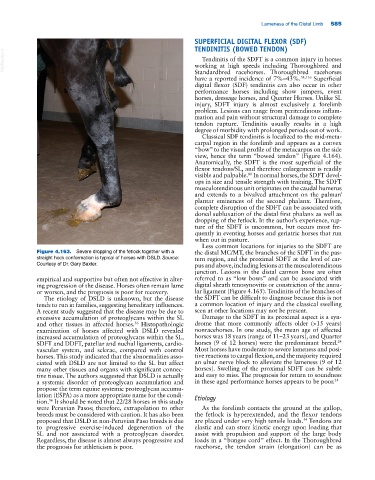
Figure 4.163. Severe dropping of the fetlock together with a the distal MC/MT, the branches of the SDFT in the pas-
straight hock conformation is typical of horses with DSLD. Source: tern region, and the proximal SDFT at the level of car-
Courtesy of Dr. Gary Baxter. pus and above, including lesions at the musculotendinous
junction. Lesions in the distal cannon bone are often
empirical and supportive but often not effective in alter- referred to as “low bows” and can be associated with
ing progression of the disease. Horses often remain lame digital sheath tenosynovitis or constriction of the annu-
or worsen, and the prognosis is poor for recovery. lar ligament (Figure 4.165). Tendinitis of the branches of
The etiology of DSLD is unknown, but the disease the SDFT can be difficult to diagnose because this is not
tends to run in families, suggesting hereditary influences. a common location of injury and the classical swelling
A recent study suggested that the disease may be due to seen at other locations may not be present.
excessive accumulation of proteoglycans within the SL Damage to the SDFT in its proximal aspect is a syn-
and other tissues in affected horses. Histopathologic drome that more commonly affects older (>15 years)
56
examination of horses affected with DSLD revealed nonracehorses. In one study, the mean age of affected
increased accumulation of proteoglycans within the SL, horses was 18 years (range of 11–23 years), and Quarter
24
SDFT and DDFT, patellar and nuchal ligaments, cardio- horses (9 of 12 horses) were the predominant breed.
vascular system, and sclerae, compared with control Most horses have moderate to severe lameness and posi-
horses. This study indicated that the abnormalities asso- tive reactions to carpal flexion, and the majority required
ciated with DSLD are not limited to the SL but affect an ulnar nerve block to alleviate the lameness (9 of 12
many other tissues and organs with significant connec- horses). Swelling of the proximal SDFT can be subtle
tive tissue. The authors suggested that DSLD is actually and easy to miss. The prognosis for return to soundness
a systemic disorder of proteoglycan accumulation and in these aged performance horses appears to be poor. 24
propose the term equine systemic proteoglycan accumu-
lation (ESPA) as a more appropriate name for the condi- Etiology
tion. It should be noted that 22/28 horses in this study
56
were Peruvian Pasos; therefore, extrapolation to other As the forelimb contacts the ground at the gallop,
breeds must be considered with caution. It has also been the fetlock is hyperextended, and the flexor tendons
54
proposed that DSLD in non‐Peruvian Paso breeds is due are placed under very high tensile loads. Tendons are
to progressive exercise‐induced degeneration of the elastic and can store kinetic energy upon loading that
SL and not associated with a proteoglycan disorder. assist with propulsion and support of the large body
Regardless, the disease is almost always progressive and loads in a “bungee cord” effect. In the Thoroughbred
the prognosis for athleticism is poor. racehorse, the tendon strain (elongation) can be as