
Page 530 - Fluid, Electrolyte, and Acid-Base Disorders in Small Animal Practice
P. 530
518 FLUID THERAPY
A
Alveolus I.S.
L
Pc
V
πc
Pi
πi
Tb
Capillary
Alveolus
I.S.
V
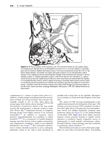
Figure 21-2 Microcirculation of the respiratory unit. The arteriole branches into the capillary plexus
surrounding alveoli. Starling's forces controlling fluid movement into or out of the capillary or interstitial
space (IS) are indicated. Capillary hydrostatic pressure in the lung must generally exceed 20 to 25 mm Hg
before edema develops. Chronically, even higher hydrostatic pressures can be tolerated before edema
develops. This is explained by the increased lymphatic drainage of the interstitium that develops in chronic
edematous states. P c , Capillary hydrostatic pressure, which forces fluid into the interstitium; p c , capillary
colloid osmotic pressure principally because of albumin, which causes fluid to be retained within the capillary;
P i , interstitial hydrostatic pressure, which is negative in the lung; p i , interstitial colloid osmotic pressure, which
is controlled by pulmonary lymphatics and maintains the interstitium relatively free of albumin; A, arteriole; V,
venule; L, lymphatic vessel; Tb, terminal bronchiole. (From Ware WW, Bonagura JD. Pulmonary edema. In:
Fox PR, editor. Canine and feline cardiology. Philadelphia: WB Saunders, 1999: 252. Medical illustration by
Felicia Paras.)
compartment (i.e., edema) or serous body cavities (i.e., perihilar and in lung lobes on the rightside, although it
effusion). A safety margin normally prevents this accumu- can accumulate in cranial and ventral regions at the same
lation of fluid, and venous pressures must increase sub- time.
stantially (usually to two or three times above the The edema of CHF develops predominantly in the
60,157,165,180
normal upper limit) before edema develops. capillary beds drained by the failing side of the heart. This
Development of pulmonary edema in the dog usually finding is pertinent because CHF is classified clinically as
requires left atrial pressure to increase acutely to more left-sided, right-sided, or biventricular. Increased pulmo-
than 20 mm Hg. 60 Substantial increases in lymphatic nary venous and capillary hydrostatic pressures cause
drainage permit much higher pressure to be tolerated pulmonary edema (see Figure 21-2), the cardinal finding
chronically. 16,34 In addition to increased venous of left-sided CHF. Right-sided heart failure increases
pressures, hypoalbuminemia can contribute to edema systemic venous pressures leading to jugular venous
formation. 60,183 As a consequence of variable lymphatic distention or pulsation, hepatic congestion, ascites, or
drainage and other factors, such as capillary permeability (infrequently in small animals) subcutaneous edema.
and compartment compliance, edema is not uniformly Increased systemic venous pressure even may contribute
distributed in the tissues. 180 This nonuniform distribu- to pulmonary edema formation. 103
tion is evident clinically inasmuch as acute cardiogenic Pleural effusions develop as a result of left-sided, right-
pulmonary edema in the dog is most prominent in sided, or, most often, biventricular failure. This finding