Page 167 - Basic Monitoring in Canine and Feline Emergency Patients
P. 167
D3 converted  to 25-
                                                                                   1-α
               Vitamin          Absorbed GI tract     hydroxycholecalciferol    hydroxylase  (–)  ↑ P
                 D3
  VetBooks.ir  (dietary)                                   [in liver]           [in kidney]
                                                                FGF-23  (–)
                                                       (–)               25-hydroxycholecalciferol
                         ↑  Parathyroid                                   converted to Calcitriol
                           hormone                                 (–)
                                                    Release Ca (and       [active form vitamin D]
                                                   phosphorus) from                             (–)
                                                        bone
                                                                           Absorbs Ca
                                                                              (and
                                                                           phosphorus)
                                                   (–)
                 ↓  Serum        Increase reabsorption Ca (and excretion     from GI
                  calcium           phosphorus) [kidney tubule]               tract
                   levels
                                                                       ↑  Serum calcium
                                                                            levels


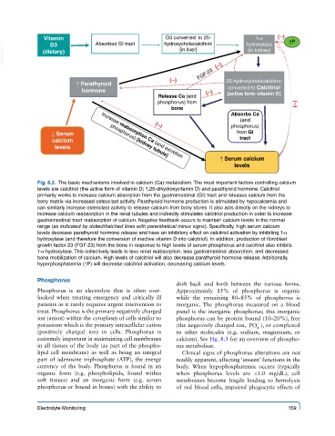
             Fig. 8.2.  The basic mechanisms involved in calcium (Ca) metabolism. The most important factors controlling calcium
             levels are calcitriol (the active form of vitamin D; 1,25-dihydroxyvitamin D) and parathyroid hormone. Calcitriol
             primarily works to increase calcium absorption from the gastrointestinal (GI) tract and releases calcium from the
             bony matrix via increased osteoclast activity. Parathyroid hormone production is stimulated by hypocalcemia and
             can similarly increase osteoclast activity to release calcium from bony stores. It also acts directly on the kidneys to
             increase calcium reabsorption in the renal tubules and indirectly stimulates calcitriol production in order to increase
             gastrointestinal tract reabsorption of calcium. Negative feedback occurs to maintain calcium levels in the normal
             range (as indicated by dotted/hatched lines with parenthetical minus signs). Specifically, high serum calcium
             levels decrease parathyroid hormone release and have an inhibitory effect on calcitriol activation by inhibiting 1-α
             hydroxylase (and therefore the conversion of inactive vitamin D into calcitriol). In addition, production of fibroblast
             growth factor 23 (FGF-23) from the bone in response to high levels of serum phosphorus and calcitriol also inhibits
             1-α hydroxylase. This collectively leads to less renal reabsorption, less gastrointestinal absorption, and decreased
             bone mobilization of calcium. High levels of calcitriol will also decrease parathyroid hormone release. Additionally,
             hyperphosphatemia (↑P) will decrease calcitriol activation, decreasing calcium levels.

             Phosphorus
                                                         shift  back  and forth  between  the various  forms.
             Phosphorus is an electrolyte that is often over-  Approximately 15% of phosphorus is organic
             looked when treating emergency and critically ill   while the remaining 80–85% of phosphorus is
             patients as it rarely requires urgent intervention to   inorganic.  The phosphorus measured on a blood
             treat. Phosphorus is the primary negatively charged   panel is the inorganic phosphorus; this inorganic
             ion (anion) within the cytoplasm of cells similar to   phosphorus can be protein bound (10–20%), free
                                                                                   −
             potassium which is the primary intracellular cation   (the negatively charged ion,  PO ), or complexed
                                                                                   4
             (positively charged ion) in cells. Phosphorus is   to  other molecules (e.g. sodium, magnesium, or
             extremely important in maintaining cell membranes   calcium). See Fig. 8.3 for an overview of phospho-
             in all tissues of the body (as part of the phospho-  rus metabolism.
             lipid cell membrane) as well as being an integral   Clinical signs of phosphorus alterations are not
             part of adenosine triphosphate (ATP), the energy   readily apparent, affecting ‘unseen’ functions in the
             currency of the body. Phosphorus is found in an   body.  When hypophosphatemia occurs (typically
             organic form (e.g. phospholipids, found within   when  phosphorus  levels  are  ≤1.0  mg/dL),  cell
             soft  tissues)  and  an inorganic  form (e.g. serum   membranes become fragile leading to hemolysis
             phosphorus or bound in bones) with the ability to   of  red  blood cells, impaired phagocytic effects of


             Electrolyte Monitoring                                                          159

