
Page 172 - Basic Monitoring in Canine and Feline Emergency Patients
P. 172
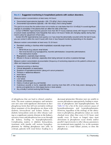
VetBooks.ir  Box 8.1.  Suggested monitoring in hospitalized patients with sodium disorders.
               Measure sodium concentrations at least every 4-6 hours:


                ● ● Documented hyponatremia (typically < 90–100 mEq/L) that is being treated
               ● ● ● ● Documented hypernatremia (typically >160–170 mEq/L) that is being treated
               The goal is to ensure that the sodium does not normalize at a rate faster than 0.5–1.0 mEq/L/h to avoid significant
               changes in osmolality in the plasma that can affect the cells of the brain.
                Correct CHRONIC hypernatremia (present more than 48–72 hours) relatively slowly with frequent monitoring
               of sodium levels (sometimes more frequently than every 4–6 hours if levels are changing rapidly) during treat-
               ment to allow for adjustment of fluid rates.
                Correct ACUTE hypernatremia more quickly (i.e. if you know the abnormality occurred within the last 8 hours,
               you can correct it within the next 8 hours) with more or less frequent monitoring depending on the situation.
               Measure sodium concentration at least every 12–24 hours:
               ● ● ● Persistent vomiting or diarrhea while hospitalized, especially large volumes
               ● ● ● Diuresis
                 # ●● Renal failure (e.g. polyuric renal failure)
                 # ●● Non-renal diuresis (e.g hyperglycemia, mannitol administration, furosemide administration)
                 # ●● Post-obstructive diuresis
               ● ● ● Regurgitation, especially large volumes
               ● ● ● Effusion in thorax or abdomen, especially when being removed via active or passive drainage
               Measure sodium concentration at presentation (frequency of rechecking depends on the patient’s clinical con-
               dition and response to treatment):
               ● ● ● Historical vomiting or diarrhea
               ● ● ● Clinical dehydration on examination
               ● ● ● Suspicion of hypoadrenocorticism (along with serum potassium)
               ● ● ● Thoracic or abdominal effusions
               ● ● ● Heart failure
               ● ● ● Renal failure
               ● ● ● Liver failure
               ● ● ● Diuretic administration
               ● ● ● History of sodium gain (e.g. salt toxicity)
               ● ● ● Cutaneous wounds or burns, especially when covering more than 20% of the body and/or damaging the
                 dermis and epidermis (i.e. 2nd degree burns or more severe)
               ● ● ● Any critically ill animal receiving fluid therapy


            of phosphorus that can lead to clinical complica-  decreased glomerular filtration rate) are unable to
            tions. The most common emergency and intensive   excrete phosphorus appropriately, leading to reten-
            care unit cases with significant alterations in their   tion of phosphorus and hyperphosphatemia. See
            phosphorus levels that will not normalize without   Table 8.3 for a list of the most common causes of
            direct treatment of the phosphorus are those with   hypophosphatemia and hyperphosphatemia.
            diabetic ketoacidosis and kidney failure. In   However, regardless of the etiology of the altera-
            diabetic ketoacidosis (and any other disease lead-  tions in phosphorus, phosphorus values are rarely
            ing to a significant osmotic diuresis in the kidney),   checked more often than every 6–12 hours (and
            excessive water and phosphorus loss occur through   usually every 24 hours or longer) since most
            the kidney, leading to hypophosphatemia.     alterations in phosphorus occur slowly and/or
            Complicating  matters  in  diabetic  ketoacidosis,   parallel improvements in other disease parameters
            treatment with insulin will worsen hypophos-  are monitored more regularly (i.e. as the azotemia
            phatemia by shifting phosphorus into cells. In   improves in a renal failure patient, so does the
            contrast, patients with renal failure (leading to a   phosphorus).


             164                                                                     E.J. Thomovsky