
Page 419 - Feline diagnostic imaging
P. 419
25.6 Hyperaldosteronism or Conn Disease 429
(a) (b)
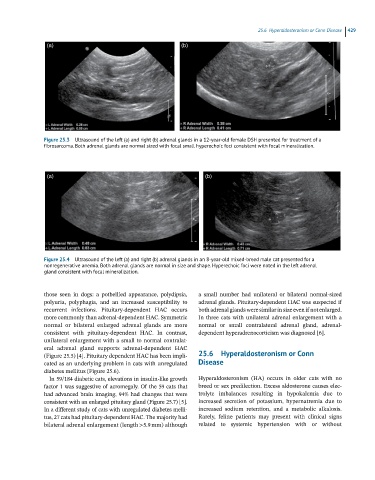
Figure 25.3 Ultrasound of the left (a) and right (b) adrenal glands in a 12-year-old female DSH presented for treatment of a
fibrosarcoma. Both adrenal glands are normal sized with focal small hyperechoic foci consistent with focal mineralization.
(a) (b)
Figure 25.4 Ultrasound of the left (a) and right (b) adrenal glands in an 8-year-old mixed-breed male cat presented for a
nonregenerative anemia. Both adrenal glands are normal in size and shape. Hyperechoic foci were noted in the left adrenal
gland consistent with focal mineralization.
those seen in dogs: a potbellied appearance, polydipsia, a small number had unilateral or bilateral normal‐sized
polyuria, polyphagia, and an increased susceptibility to adrenal glands. Pituitary‐dependent HAC was suspected if
recurrent infections. Pituitary‐dependent HAC occurs both adrenal glands were similar in size even if not enlarged.
more commonly than adrenal‐dependent HAC. Symmetric In three cats with unilateral adrenal enlargement with a
normal or bilateral enlarged adrenal glands are more normal or small contralateral adrenal gland, adrenal‐
consistent with pituitary‐dependent HAC. In contrast, dependent hyperadrenocorticism was diagnosed [6].
unilateral enlargement with a small to normal contralat-
eral adrenal gland supports adrenal‐dependent HAC
(Figure 25.5) [4]. Pituitary dependent HAC has been impli- 25.6 Hyperaldosteronism or Conn
cated as an underlying problem in cats with unregulated Disease
diabetes mellitus (Figure 25.6).
In 59/184 diabetic cats, elevations in insulin‐like growth Hyperaldosteronism (HA) occurs in older cats with no
factor 1 was suggestive of acromegaly. Of the 59 cats that breed or sex predilection. Excess aldosterone causes elec-
had advanced brain imaging, 94% had changes that were trolyte imbalances resulting in hypokalemia due to
consistent with an enlarged pituitary gland (Figure 25.7) [5]. increased secretion of potassium, hypernatremia due to
In a different study of cats with unregulated diabetes melli- increased sodium retention, and a metabolic alkalosis.
tus, 27 cats had pituitary‐dependent HAC. The majority had Rarely, feline patients may present with clinical signs
bilateral adrenal enlargement (length >5.9 mm) although related to systemic hypertension with or without