
Page 414 - Feline diagnostic imaging
P. 414
24.6 Diseisi of tsf eancsei 423
24.6.5 Interventional Procedures
Fine needle aspiration of the pancreas or tissue surround-
ing the pancreas has been done with ultrasound guidance
to assist in the diagnosis of pancreatitis or pancreatic
masses (Figure 24.38). The gold standard for diagnosis of
pancreatitis is histopathologic sampling of the pancreas;
however, a single sample may miss the diagnosis of pan-
creatitis due to the patchy distribution of pancreatic
change associated with pancreatitis. In general, surgical
pancreatic biopsy is reserved for cases with obstruction of
the CBD [7].
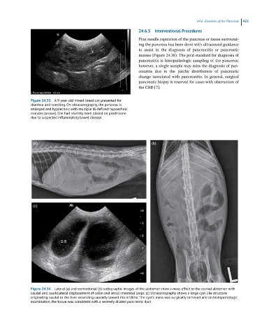
Figure 24.33 A 9-year-old mixed-breed cat presented for
diarrhea and vomiting. On ultrasonography, the pancreas is
enlarged and hypoechoic with multiple ill-defined hypoechoic
nodules (arrows). She had recently been placed on prednisone
due to suspected inflammatory bowel disease.
(a) (b)
(c)
Figure 24.34 Lateral (a) and ventrodorsal (b) radiographic images of the abdomen show a mass effect in the cranial abdomen with
caudal and caudolateral displacement of colon and small intestinal loops. (c) Ultrasonography shows a large cyst-like structure
originating caudal to the liver extending caudally toward the midline. The cystic mass was surgically removed and on histopathologic
examination, the tissue was consistent with a severely dilated pancreatic duct.