
Page 411 - Feline diagnostic imaging
P. 411
420 24 Pancreas
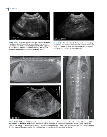
Figure 24.25 A 15-year-old diabetic Maine Coon presented for Figure 24.26 A 6-year-old male DSH presented for vomiting,
vomiting and weight loss. A large hypoechoic mass is present inappetence, and urinary incontinence. The pancreas is enlarged
associated with the duodenum and pylorus. The mass appears in and mildly hypoechoic. Fine needle aspiration of the pancreas
the location for the pancreas. Due to the concurrent regional and free abdominal fluid revealed carcinoma.
lymphadenopathy, pancreatic carcinoma was suspected.
(a) (b)
(c)
Figure 24.27 A 19-year-old DSH presented for vomiting and diarrhea. A soft tissue mass is noted in the cranial abdomen caudal to
the stomach on the lateral image (a) and in a similar location toward the midline on the ventrodorsal image (b). Both kidneys are
smaller than normal. On abdominal ultrasound, a heterechoic mass with focal cavitations is seen with reactive omentum surrounding
it in the region of the pancreas (c). A fine needle aspirate was consistent with pancreatic carcinoma.