
Page 413 - Feline diagnostic imaging
P. 413
422 24 Pancreas
(a) (b)
(c)
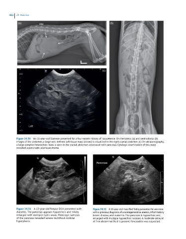
Figure 24.30 An 11-year-old Siamese presented for a four-month history of inappetence. On the lateral (a) and ventrodorsal (b)
images of the abdomen, a large well-defined soft tissue mass (arrows) is visualized in the right cranial abdomen. (c) On ultrasonography,
a large complex heterechoic mass is seen in the cranial abdomen associated with pancreas. Cytologic examination of this mass
revealed a pancreatic adenocarcinoma.
Figure 24.31 A 13-year-old female DSH presented with Figure 24.32 A 10-year-old male Red Tabby presented for anorexia
diabetes. The pancreas appears hypoechoic and mildly with a previous diagnosis of a nonregenerative anemia, inflammatory
enlarged with multiple cystic areas. Histologic samples bowel disease, and leukemia. The pancreas is hypoechoic and
of the pancreas revealed severe multifocal nodular enlarged with multiple hypoechoic nodules. A moderate amount
hyperplasia. of free abdominal fluid is present. Pancreatitis was suspected.