
Page 433 - Feline diagnostic imaging
P. 433
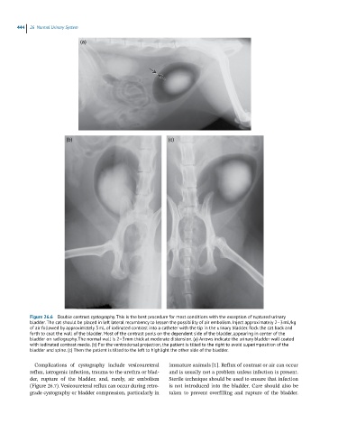
444  26  Normal Urinary System

                              (a)


                         (b)                                  (c)


            Figure 26.6  Double contrast cystography. This is the best procedure for most conditions with the exception of ruptured urinary
            bladder. The cat should be placed in left lateral recumbency to lessen the possibility of air embolism. Inject approximately 2–3 mL/kg
            of air followed by approximately 3 mL of iodinated contrast into a catheter with the tip in the urinary bladder. Rock the cat back and
            forth to coat the wall of the bladder. Most of the contrast pools on the dependent side of the bladder, appearing in center of the
            bladder on radiography. The normal wall is 2–3 mm thick at moderate distension. (a) Arrows indicate the urinary bladder wall coated
            with iodinated contrast media. (b) For the ventrodorsal projection, the patient is tilted to the right to avoid superimposition of the
            bladder and spine. (c) Then the patient is tilted to the left to highlight the other side of the bladder.


              Complications  of  cystography  include  vesicoureteral   immature animals [1]. Reflux of contrast or air can occur
            reflux, iatrogenic infection, trauma to the urethra or blad-  and is usually not a problem unless infection is present.
            der,  rupture  of  the  bladder,  and,  rarely,  air  embolism   Sterile technique should be used to ensure that infection
            (Figure 26.7). Vesicoureteral reflux can occur during retro-  is not introduced into the bladder. Care should also be
            grade cystography or bladder compression, particularly in   taken  to  prevent  overfilling  and  rupture  of  the  bladder.