
Page 501 - Feline diagnostic imaging
P. 501
29.5 DesheMeshees of Thef mphehea 513
(a) (b)
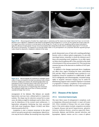
Figure 29.14 Ultrasonography of medial iliac lymph nodes in lymphoma. (a) The medial iliac lymph node of an 8-year-old domestic
shorthair with multicentric lymphoma is decreased in echogenicity and enlarged at 1.7 × 0.5 cm. (b) The medial iliac lymph nodes can
be imaged from either the lateral or ventral aspect. In this image of a 19-year-old cat with lymphoma (diffuse large centroblastic
type), the ultrasound beam passes through the urinary bladder (B) to image the node which is rounded, measuring 0.7 × 0.9 cm and
decreased in echogenicity. The appearance of enlarged lymph nodes is not pathognomonic for lymphoma. Abnormal-appearing lymph
nodes should be aspirated or biopsied to determine the cause.
poorly demarcated areas of lysis with resulting pathologic
fracture with or without spinal compression. In cats with
neurologic lesions, remember to check the kidneys to see if
there was preexisting renal lymphoma. In an older report
on feline renal lymphoma 40% of the cats died as the result
of involvement of the nervous system [16]. This may be less
common because of the decrease in numbers of FeLV‐posi-
tive cats [17].
In addition to the changes mentioned above, lymphoma
can affect other organs including the brain, pericardium
[18], and skin. When a retrobulbar mass is present in a cat,
lymphoma should be considered as a differential. In addi-
tion to radiography and ultrasonography, computed tomog-
Figure 29.15 Ultrasonography of lymphoma in mesenteric lymph
nodes. A 9-year-old domestic shorthair was admitted with a history raphy or magnetic resonance imaging may be useful for
of chronic vomiting and anorexia. On ultrasonography, there was determining prognosis and to monitor response to treatment
enlargement of the mesenteric lymph nodes and a focal area of or progression of disease. Lymphoma can be multicentric
thickened intestinal wall. Multicentric lymphoma (B cell and may have a secondary effect on other organs [11].
phenotype) was diagnosed on histopathology after euthanasia.
The mesenteric lymph node shown here is rounded, and more
rounded and larger than normal.
29.5 Diseases of the Spleen
enlargement of the kidneys. The kidneys are usually
hyperechoic and may have a dilated pelvis or hypoechoic 29.5.1 Generalized Splenomegaly
nodules. The shape may be relatively normal but in some
cases, the kidneys might be misshapen and in others, there The most likely cause of enlargement is neoplasia, such
may be disturbance of the normal renal architecture. A as lymphoma (discussed previously) or mast cell tumor
hypoechoic subcapsular thickening has been associated [10]. Other examples of disease that can cause enlarge-
with both renal lymphoma (Figure 29.22d,f) and feline ment of the entire spleen include congestion secondary
infectious peritonitis [15]. to hepatic disease and portal hypertension, splenitis,
Lymphoma is also a common neoplasm in the spinal immune‐mediated disease, parasites especially Hemo
cord of cats and is second only to meningioma. Spinal inva- bartonella, Babesia, and Erlichia, mycotic infection, and
sion may be focal or diffuse. Surrounding bone may exhibit hyperplasia. Splenic torsion can be a cause of splenic