
Page 502 - Feline diagnostic imaging
P. 502
514 29 Hemolymphatic System
(a) (b)
(c)
(d)
(e)
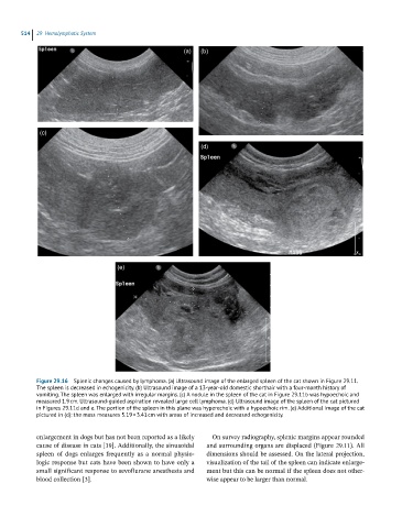
Figure 29.16 Splenic changes caused by lymphoma. (a) Ultrasound image of the enlarged spleen of the cat shown in Figure 29.11.
The spleen is decreased in echogenicity. (b) Ultrasound image of a 13-year-old domestic shorthair with a four-month history of
vomiting. The spleen was enlarged with irregular margins. (c) A nodule in the spleen of the cat in Figure 29.11b was hypoechoic and
measured 1.9 cm. Ultrasound-guided aspiration revealed large cell lymphoma. (d) Ultrasound image of the spleen of the cat pictured
in Figures 29.11d and e. The portion of the spleen in this plane was hyperechoic with a hypoechoic rim. (e) Additional image of the cat
pictured in (d): the mass measures 5.19 × 3.41 cm with areas of increased and decreased echogenicity.
enlargement in dogs but has not been reported as a likely On survey radiography, splenic margins appear rounded
cause of disease in cats [19]. Additionally, the sinusoidal and surrounding organs are displaced (Figure 29.11). All
spleen of dogs enlarges frequently as a normal physio- dimensions should be assessed. On the lateral projection,
logic response but cats have been shown to have only a visualization of the tail of the spleen can indicate enlarge-
small significant response to sevoflurane anesthesia and ment but this can be normal if the spleen does not other-
blood collection [3]. wise appear to be larger than normal.