
Page 1693 - Cote clinical veterinary advisor dogs and cats 4th
P. 1693
851.e2 Pyloric Outflow Obstruction
Pyloric Outflow Obstruction Client Education
Sheet
VetBooks.ir
be reduced if patient is fed small, frequent meals
BASIC INFORMATION
rowed outflow path of pylorus (described as
of a liquid diet. • Fluoroscopy with contrast: highlights nar-
Definition a beak sign) and ineffective emptying
Syndrome arising from many possible pathologic PHYSICAL EXAM FINDINGS • Abdominal ultrasound: increased mucosal or
processes (congenital or acquired) that impair Failure to gain weight as adolescents and poor muscular thickness of the pylorus; depends
movement of ingesta from the stomach to the body condition scores as adults are possible. on obtaining clear transverse image
duodenum due to obstruction or narrowing of • Gastroscopy (p. 1098): narrowed, tight,
the pyloric lumen. Etiology and Pathophysiology pylorus or thick mucosal folds around or
• Congenital: circular smooth muscle hyper- covering the pylorus forming a hoodlike
Synonyms trophy (type 1), combination of circular appearance; skilled endoscopists may find
Antral pyloric obstruction, pyloric stenosis, smooth muscle hypertrophy and mucosal passing the endoscope through the pylorus
gastric outlet obstruction, hypertrophic pyloric hyperplasia (type 2), or primarily mucosal difficult. Histopathology of endoscopic
gastropathy hyperplasia (type 3) contribute to functional biopsies may or may not reveal mucosal
obstruction of the pylorus. Cause is unknown hypertrophy.
Epidemiology and inheritance not determined. • Surgery, histopathology: may be needed to
SPECIES, AGE, SEX • Acquired confirm diagnosis by probing the characteris-
Dogs, and rarely cats, of either sex and any ○ Pyloric obstruction secondary to mucosal tic narrowed pyloric lumen. Histopathology
age can be affected. Congenital disease may be hypertrophy characterized histologically features as described (see Etiology and Patho-
seen in puppies and kittens to young adults. by mucosal foveolar and glandular physiology above); superficial erosions from
hyperplasia, cystic glandular dilatation, concurrent inflammation and mechanical
GENETICS, BREED PREDISPOSITION superficial mucosal ulcerations, and trauma can also be present.
Congenital pyloric outflow obstruction occurs minimal cellular infiltrates; muscular layer
more often in small-breed dogs but can occur in may be minimally involved TREATMENT
any purebred or mix-breed dog. The pattern of ○ Accentuated rugal folds contribute to
inheritance and identification of genetic markers obstruction. Treatment Overview
are not reported. Brachycephalic breeds (Boston Treatment goal is to alleviate pyloric obstruction
terrier, boxer, French bulldog, and bulldog DIAGNOSIS when possible.
varieties) are more commonly affected. Other
breeds at risk include Maltese, Lhasa apso, shih Diagnostic Overview Acute General Treatment
tzu, Pekingese, poodles, and rottweiler. Diagnosis hinges on ruling out other causes Correct fluid deficits and electrolyte abnormali-
of chronic vomiting and demonstration of ties if present.
RISK FACTORS impaired gastric emptying.
Chronic gastrointestinal disease (see Associated Chronic Treatment
Disorders below) can be a risk factor for the Differential Diagnosis Surgical interventions such as pyloroplasty tech-
acquired form. • Esophageal disease (for patients that seem niques, gastroduodenostomy, or pyloromyotomy
to be regurgitating) are often required to alleviate clinical signs,
ASSOCIATED DISORDERS • Foreign body particularly for the congenital form.
Any concurrent chronic, irritating, or inflam- • Mucosal polyps
matory conditions (inflammatory bowel disease, • Chronic gastritis
chronic gastritis, gastric infection, foreign • Helicobacter spp
bodies, gastric parasites) may be a risk factor. • Gastric neoplasia
• Physaloptera spp
Clinical Presentation • Trichobezoar
DISEASE FORMS/SUBTYPES • Gastrinoma and parietal cell hyperplasia
• Congenital form is often called pyloric stenosis. • Giant hypertrophic gastritis (Menetrier-like
• Acquired form, with descriptions such as disease) ± carcinoma (rare)
pyloric mucosal hypertrophy, develops as
response to a concurrent disease or from Initial Database
undetermined cause. Intraluminal lesions • CBC, biochemical profile, urinalysis: rule
(e.g., polyps, tumors, foreign bodies, out systemic causes of chronic vomiting;
parasites) or extraluminal compression (e.g., hypokalemia, hypochloremia, and acid-base
gastric serosal tumor, adjacent pancreatic imbalances may occur.
cancer) may also cause clinical signs. • Fecal examinations
• Abdominal radiographs: rule out obvious
HISTORY, CHIEF COMPLAINT foreign bodies
Patients are often presented for intermittent • Thoracic radiographs: rule out obvious
to persistent vomiting that can be projectile. esophageal disease in patients that appear
Vomiting, often of undigested food, may to be regurgitating
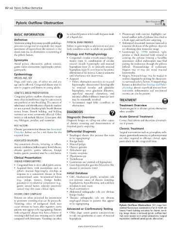
occur minutes to hours after ingestion; some Pyloric Outflow Obstruction Still image from
contrast fluoroscopic examination of an 8-month old
patients may appear to be regurgitating. Patients Advanced or Confirmatory Testing female French bulldog evaluated for chronic vomit-
with congenital disease may have a history of • Older dogs: serum gastrin concentrations ing. Image shows a narrowed pyloric outflow tract
vomiting solid food since weaning and be small to rule out gastrinoma as cause of mucosal that never opened with antral contractions. Surgical
compared with littermates. Vomiting can often hyperplasia exploration confirmed antral pyloric obstruction.
www.ExpertConsult.com