
Page 1695 - Cote clinical veterinary advisor dogs and cats 4th
P. 1695
852 Pyoderma
CONTAGION AND ZOONOSIS dermatitis with superficial ulceration but follicle, rupture of the follicle (furuncles)
Staphylococcus pseudintermedius, the most also a component of deep folliculitis and occurs. This leads to a pyogranulomatous
VetBooks.ir sidered to be rarely pathogenic to humans. may be thickened and plaquelike and have of the host. This reaction occurs initially in
endogenous foreign-body reaction on the part
occasional furunculosis. Clinically, this lesion
common cause of canine pyoderma, is con-
satellite papules and pustules.
the dermis and is an inflammatory response
GEOGRAPHY AND SEASONALITY
a papular/pustular eruption on the bridge
Warm and humid environments predispose • Nasal folliculitis and furunculosis: initially to keratin, bacterial organisms, and cellular
debris.
animals to pyoderma. of the nose that progresses to ulceration, • More than 90% of cases in dogs are caused
crusting, and hemorrhage by coagulase-positive S. pseudintermedius, a
Clinical Presentation • Pedal furunculosis (bacterial pododermatitis): normal component of canine skin flora. A
DISEASE FORMS/SUBTYPES interdigital papulonodules, hemorrhagic small number of cases are caused by Staphy-
Surface pyoderma: infection restricted to the bullae, and ulceration with draining tracts lococcus aureus, the most common human
surface layer of the epidermis: and fibrosis; alopecia secondary to licking pathogen. Other staphylococcal bacteria (e.g.,
• Dog: intertrigo (skinfold pyoderma), acute • Callus pyoderma: develops over pressure the coagulase-positive Staphylococcus schleiferi
moist dermatitis (pyotraumatic dermatitis, points. Skin is thickened, fibrotic, and subsp coagulans and the coagulase-negative
hot spots), BOGS hyperpigmented with foci of papules, Staphylococcus schleiferi subsp schleiferi) are
Superficial pyoderma: infection involving the pustules, and ulceration. less commonly associated with bacterial
epidermis and the infundibular portion of the • Postgrooming folliculitis/furunculosis: pyoderma.
hair follicle: papular/pustular rash, crust formation, ○ Staphylococcus intermedius was reclassified
• Dog: impetigo, SBF, exfoliative superficial self-induced alopecia as S. pseudintermedius in 2007.
pyoderma (ESP, superficial spreading pyo- • German shepherd deep pyoderma: lesions • The gram-negative organisms, Proteus spp,
derma), mucocutaneous pyoderma include ulcerations and draining tracts on Escherichia coli, and Pseudomonas spp, may
Deep pyoderma: bacterial infection extending the lateral thighs, trunk, and groin ± lips. act as secondary invaders. Deep pyoderma
beyond the hair follicle to involve the dermis • Infected feline acne: comedones, papules, is occasionally associated with Actinomyces,
and subcutis, which may lead to cellulitis: pustules, and alopecia confined to the Nocardia, mycobacteria, and Actinobacillus
• Dog: acne, pyotraumatic furunculosis, nasal mandibular and perilabial areas spp.
folliculitis (inflammation of the hair follicle) • Feline bite wounds/abscess/cellulitis: swell- • In cats, Pasteurella multocida and beta-
and furunculosis (simultaneous occurrence ing, pain, and alopecia in the affected area; hemolytic Streptococcus are routinely involved.
of many furuncles, which are inflamed hair possible dermal and cutaneous necrosis with
follicles that have ruptured, triggering pyo- ulceration, purulent exudate, and hemorrhage DIAGNOSIS
granulomatous dermal inflammation), inter-
digital furunculosis/pododermatitis, infected Etiology and Pathophysiology Diagnostic Overview
acral lick dermatitis (lick granuloma), callus • Canine skin is characterized by a relatively The dermatologic exam alone may provide a
pyoderma, and postgrooming furunculosis; thin stratum corneum, a paucity of intercel- strong suspicion of pyoderma (see signs listed
German shepherd: deep pyoderma lular lipids, lack of a follicular plug, and a above). With clinical signs of deep pyoderma,
• Cat: bite wounds, cellulitis, abscess, feline higher pH than that of humans and other systemic signs, or recurrence despite therapy,
acne domestic species. This predisposes the dog to additional tests beyond basic dermatologic
overgrowth of commensal flora and coloniza- testing are often indicated to rule out predispos-
HISTORY, CHIEF COMPLAINT tion by potentially pathogenic bacteria. ing causes.
Animals typically are presented for evaluation • Superficial infection may occur if the integrity
of skin changes: pustules, crusts, epidermal of the skin is weakened by trauma or there Differential Diagnosis
collarettes, hair loss, and/or pruritus. Animals are changes in surface immunity. • Demodicosis
with deep pyoderma may show signs of pain. • Deep bacterial infections are generally an • Dermatophytosis
extension of a superficial pyoderma. As • Pemphigus foliaceus
PHYSICAL EXAM FINDINGS infection progresses deeper into the hair • Cutaneous epitheliotropic lymphoma
• Impetigo: small nonfollicular pustules and
crusts on the ventral abdomen
• BOGS: pruritus, greasy coat, offensive odor,
erythema, lichenification, hyperpigmenta-
tion, excoriations, and alopecia
• SBF: papules, pustules, and epidermal col-
larettes with patchy alopecia, producing a
moth-eaten appearance of the haircoat over
the trunk. Resolving lesions may show central
hyperpigmentation (bull’s-eye lesion).
• ESP: large epidermal collarettes with an
erythematous leading edge noted over the
trunk; associated exudate may form crusts.
• Canine acne: deep folliculitis and furuncu-
losis, with crusting and possible scarring on
the lips and chin of young dogs
• Mucocutaneous pyoderma: crusts and ero-
sions affecting the lip margin, nasal planum,
eyelid margin, vulva, prepuce, and perianal
area
• Pyotraumatic folliculitis and furunculosis:
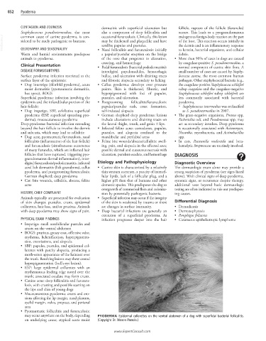
may occur anywhere on the body, depending PYODERMA Epidermal collarettes on the ventral abdomen of a dog with superficial bacterial folliculitis.
on underlying cause; atypical acute moist (Copyright Dr. Manon Paradis.)
www.ExpertConsult.com