
Page 1776 - Cote clinical veterinary advisor dogs and cats 4th
P. 1776
891.e2 Right Bundle Branch Block
Right Bundle Branch Block
VetBooks.ir Etiology and Pathophysiology
• Cardiomyopathy
BASIC INFORMATION
• Congenital heart disease • With RBBB, impulse formation in the sino-
Definition • Heartworm disease atrial node, atrial depolarization, and passage
The intracardiac conduction disturbance Clinical Presentation of the impulse through the atrioventricular
involves failure of normal (rapid) conduction (AV) node can occur normally, but distribu-
from the bundle of His through the right DISEASE FORMS/SUBTYPES tion of the impulse through the Purkinje
bundle branch to the Purkinje fibers in the right • Incomplete or complete RBBB, depending on fibers in the right ventricle is blocked.
ventricle. The result is a wide, bizarre-appearing where the block occurs in the right bundle • Conduction to the right ventricle occurs
QRS complex on the electrocardiogram (ECG). branch but is very slow because it must travel from
Right bundle branch block (RBBB) has no • Intermittent bundle branch block (RBBB muscle cell to muscle cell rather than through
clinically meaningful hemodynamic effect on or, less commonly, left bundle branch the rapid conduction system.
patients and is often a variant of normal. Its block [LBBB]) may occur that is heart rate • This results in a marked delay in conduc-
main clinical importance is that it must not dependent. tion to the right ventricle, and the QRS
be misinterpreted as a ventricular arrhythmia. complex becomes wide and bizarre on the
HISTORY, CHIEF COMPLAINT ECG.
Epidemiology RBBB is an electrocardiographic phenomenon • This slow conduction toward the right results
SPECIES, AGE, SEX only; no overt physical manifestations are in a right-axis deviation of the QRS mean
Dogs of any age and either sex; less common expected from RBBB itself. electrical axis on the surface ECG.
in cats
PHYSICAL EXAM FINDINGS DIAGNOSIS
GENETICS, BREED PREDISPOSITION Typically, RBBB is clinically silent, and no
Incomplete RBBB has been recognized in a clinical signs are observed. However, in some Diagnostic Overview
family of beagles in association with congenital individuals, split heart sounds may occur The diagnosis is purely electrocardiographic:
right ventricular structural disease. due to prolonged right ventricular ejection it is suspected when QRS duration is longer
time and delayed closure of the tricuspid than normal; S waves are seen in leads I, II,
ASSOCIATED DISORDERS (splitting of the first heart sound) and pul- III, and aVF and the left precordial leads, and
RBBB often occurs in normal animals, but it monic (splitting of the second heart sound) the rhythm appears to be supraventricular in
can be associated with underlying structural valves. A split second heart sound is most origin. The inherent rhythm of the heart is
heart diseases: common. not affected.
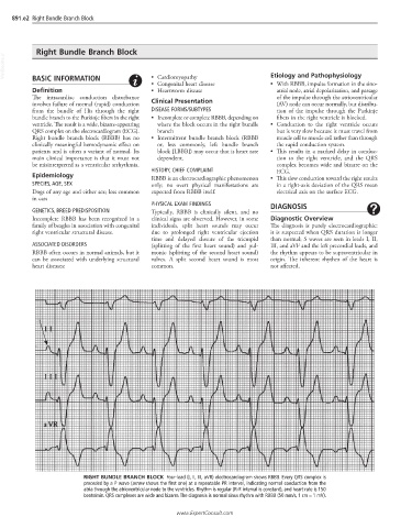
RIGHT BUNDLE BRANCH BLOCK Four-lead (I, II, III, aVR) electrocardiogram shows RBBB. Every QRS complex is
preceded by a P wave (arrow shows the first one) at a repeatable PR interval, indicating normal conduction from the
atria through the atrioventricular node to the ventricles. Rhythm is regular (R-R interval is constant), and heart rate is 150
beats/min. QRS complexes are wide and bizarre. The diagnosis is normal sinus rhythm with RBBB (50 mm/s, 1 cm = 1 mV).
www.ExpertConsult.com