
Page 1779 - Cote clinical veterinary advisor dogs and cats 4th
P. 1779
Round Cell Tumors 893
Client Education owners’ cuticles be exposed to infective tick rickettsialpox, and typhus. In Greene CE, editor:
• RMSF in dogs is a warning to owners that hemolymph/feces. Infectious diseases of the dog and cat, ed 4, St.
Louis, 2012, Saunders, pp 259-270.
VetBooks.ir • Ticks removed from pets must not be SUGGESTED READING AUTHOR: Meryl P. Littman, VMD, DACVIM Diseases and Disorders
ticks in their area carry RMSF (other dogs
and people may be exposed).
EDITOR: Joseph Taboada, DVM, DACVIM
Greene CE, et al: Rocky Mountain and Mediter-
removed by unprotected fingers, lest
ranean spotted fevers, cat-flea typhuslike illness,
Round Cell Tumors Bonus Material Client Education
Sheet
Online
BASIC INFORMATION cutaneous and systemic histiocytosis, and his- nasal/pharyngeal, mediastinal, central
tiocytic sarcoma), and melanocytic tumors. nervous system (CNS), and kidneys;
Definition Each of these is discussed in greater detail multicentric lymphoma can also occur,
• Cells can be classified based on certain in individual chapters. especially in cats with feline leukemia virus
common cytologic features: epithelial, (FeLV).
mesenchymal, and round cells. After cell Synonym ○ Solitary plasma cell tumors most com-
types are identified, additional cytologic Discrete cell tumors monly occur in dogs as skin and subcu-
criteria are used to help determine whether Epidemiology taneous masses (plasmacytomas) but have
cells are normal or part of an inflammatory also been reported in bones and the GI/
or neoplastic process. SPECIES, AGE, SEX respiratory tracts. Multiple myeloma is
• Round cells are often called discrete cells Depends on tumor type the systemic form of plasma cell cancer
because they exist individually in tissues Clinical Presentation and can affect organ systems such as the
rather than adhere to other cells or a matrix. bone marrow and lymphoid organs.
Round cell tumors are typically composed of DISEASE FORMS/SUBTYPES • MCTs (mastocytomas, mast cell sarcomas)
a homogeneous population of cells that have • Lymphoproliferative diseases ○ In dogs, the most common form is skin/
well-defined cytoplasmic margins, are round, ○ Lymphoma (malignant lymphoma, subcutaneous masses. The prognosis varies
and have a round nucleus. Exceptions to this lymphosarcoma) is seen in dogs and and depends on tumor grade, clinical
include histiocytic sarcomas (multinucleate cats of all ages and can affect any organ presentation, breed, and stage of disease.
giant cells with irregular nuclear shapes are system. Lymphocytic (small cell) and lym- ○ In cats, skin, splenic, and visceral forms
common) and melanoma (cells have variable phoblastic (large cell) forms have varied may occur.
appearances and can be round, epithelial, presentations, clinical signs, and treatment • Transmissible venereal tumors (TVT) are
or mesenchymal cells). Distinct cytologic options. In dogs, the most common form transplanted from dog to dog by direct
characteristics of each type of round cell is lymphoblastic lymphoma, affecting the contact (sniffing/licking/sexual contact). TVT
aid in definitive diagnosis. lymph nodes and lymphoid organs. In is most commonly seen in intact dogs in the
• Round cell tumor types include lympho- cats, solitary-site lymphocytic and lym- genital regions and oral and nasal cavities
proliferative diseases (e.g., lymphoma, phoblastic lymphoma can occur, affecting and might regress in dogs with a competent
plasma cell tumor, multiple myeloma), mast tissues that include gastrointestinal (GI), immune system.
cell tumor (MCT), transmissible venereal
tumor, histiocytic tumors (i.e., histiocytomas,
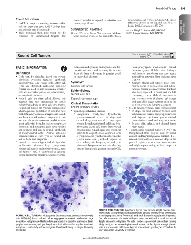
ROUND CELL TUMORS Lymphoma (lymph node aspirate, Wright-Giemsa stain).
Intermediate to large lymphoblasts predominate, admixed with few small lymphocytes,
ROUND CELL TUMORS Histiocytoma (cutaneous mass, aqueous Romanowsky many large pale-staining free nuclei, and small basophilic cytoplasmic fragments.
stain [Diff-Quik]). Round cells with a fried egg appearance contain moderate to large The cells have open chromatin with prominent nucleoli and a small amount of
amounts of cytoplasm and fairly uniform paracentral to eccentric oval nuclei. Small deeply basophilic cytoplasm. The cells contain a prominent perinuclear clearing
lymphocytes are seen, and lymphocytic or mixed inflammation may be prominent (Golgi zone), as can be seen in plasma cell tumors, but the high nuclear-cytoplasmic
in aspirates, particularly as tumors regress. (Courtesy Dr. Mary Leissinger, University ratio and chromatin pattern are typical of neoplastic lymphocytes. (Courtesy Dr.
of Florida.) Mary Leissinger, University of Florida.)
www.ExpertConsult.com