
Page 1784 - Cote clinical veterinary advisor dogs and cats 4th
P. 1784
Round Cell Tumors 894.e1
VetBooks.ir Diseases and Disorders
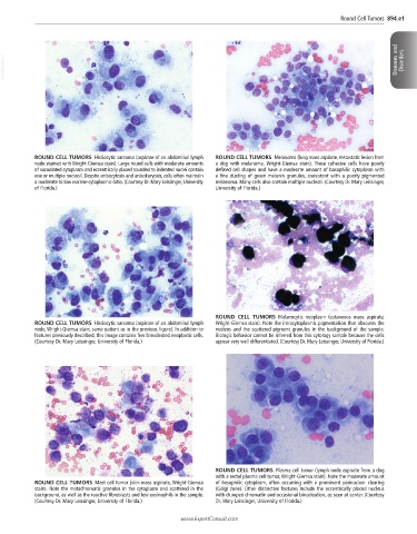
ROUND CELL TUMORS Histiocytic sarcoma (aspirate of an abdominal lymph ROUND CELL TUMORS Melanoma (lung mass aspirate, metastatic lesion from
node stained with Wright-Giemsa stain). Large round cells with moderate amounts a dog with melanoma. Wright-Giemsa stain). These cohesive cells have poorly
of vacuolated cytoplasm and eccentrically placed rounded to indented nuclei contain defined cell shapes and have a moderate amount of basophilic cytoplasm with
one or multiple nucleoli. Despite anisocytosis and anisokaryosis, cells often maintain a fine dusting of green melanin granules, consistent with a poorly pigmented
a moderate to low nuclear-cytoplasmic ratio. (Courtesy Dr. Mary Leissinger, University melanoma. Many cells also contain multiple nucleoli. (Courtesy Dr. Mary Leissinger,
of Florida.) University of Florida.)
ROUND CELL TUMORS Melanocytic neoplasm (cutaneous mass aspirate,
ROUND CELL TUMORS Histiocytic sarcoma (aspirate of an abdominal lymph Wright-Giemsa stain). Note the intracytoplasmic pigmentation that obscures the
node, Wright-Giemsa stain, same patient as in the previous figure). In addition to nucleus and the scattered pigment granules in the background of the sample.
features previously described, this image contains few binucleated neoplastic cells. Biologic behavior cannot be inferred from this cytology sample because the cells
(Courtesy Dr. Mary Leissinger, University of Florida.) appear very well differentiated. (Courtesy Dr. Mary Leissinger, University of Florida.)
ROUND CELL TUMORS Plasma cell tumor (lymph node aspirate from a dog
with a rectal plasma cell tumor, Wright-Giemsa stain). Note the moderate amount
ROUND CELL TUMORS Mast cell tumor (skin mass aspirate, Wright-Giemsa of basophilic cytoplasm, often occurring with a prominent perinuclear clearing
stain). Note the metachromatic granules in the cytoplasm and scattered in the (Golgi zone). Other distinctive features include the eccentrically placed nucleus
background, as well as the reactive fibroblasts and few eosinophils in the sample. with clumped chromatin and occasional binucleation, as seen at center. (Courtesy
(Courtesy Dr. Mary Leissinger, University of Florida.) Dr. Mary Leissinger, University of Florida.)
www.ExpertConsult.com