
Page 2028 - Cote clinical veterinary advisor dogs and cats 4th
P. 2028
1014 Urolithiasis, Oxalate
• The mortality rate is increased for animals with • Asepsis (urethral and peritoneal drainage uroabdomen often have concurrent disease or
concurrent injuries or underlying neoplasia. catheters, sterile collection system) is essential injuries and may have a long recovery period;
VetBooks.ir PEARLS & CONSIDERATIONS • In dogs and cats, lower urinary tract injuries benefits their recovery.
for preventing secondary infection.
adequate attention to analgesia and nutrition
are more common than renal/ureteral inju-
Comments
• Consider uroabdomen in animals with a ries. Contrast cystography and retrograde SUGGESTED READING
urethrography take precedence over IV
Stafford JR, et al: A clinical review of pathophysiology,
history of abdominal or pelvic trauma, urinary excretory urography. diagnosis, and treatment of uroabdomen in the dog
obstruction, or urethral catheterization. • Surgical repair should not be attempted until and cat. J Vet Emerg Crit Care 23:216-229, 2013.
• A palpable bladder and the ability to void the animal has been stabilized with fluid AUTHOR: Gareth J. Buckley, VetMB, MA, DACVECC,
urine do not rule out uroabdomen. therapy and abdominal drainage. DECVECC
• The abdominal fluid obtained is serosan- EDITOR: Benjamin M. Brainard VMD, DACVAA,
guineous and may not look like urine; Technician Tips DACVECC
creatinine and potassium concentrations of Fluid therapy is a challenge in these patients,
the fluid (compared with serum or plasma) and accurate recording of fluid administra-
are diagnostic. tion and fluid output is crucial. Patients with
Urolithiasis, Oxalate Client Education
Sheet
BASIC INFORMATION • Obesity PHYSICAL EXAM FINDINGS
• Diets designed to minimize struvite forma- Physical exam is usually unremarkable; abnor-
Definition tion in cats malities may include
Urinary tract stones (uroliths) composed of • Hematuria (stains on prepuce, vulva, or
calcium oxalate occur commonly in dogs and ASSOCIATED DISORDERS hocks)
cats. Feline lower urinary tract signs/disease • Painful urinary bladder
(FLUTS/D), urethral obstruction, chronic • Palpable cystic calculi
Synonyms kidney disease (CKD), urinary tract infection • Palpable urethral calculi (by digital rectal
• Calcium oxalate dihydrate: weddellite (UTI), nephroureterolithiasis exam in dogs)
• Calcium oxalate monohydrate: whewellite • Enlarged, turgid bladder if urethral
Clinical Presentation
obstruction
Epidemiology HISTORY, CHIEF COMPLAINT • Renomegaly if secondary hydronephrosis or
SPECIES, AGE, SEX Clinical signs may be absent or include small irregular kidneys if CKD
Dogs: • Hematuria • Findings associated with predisposing factors
• Increased frequency over the past 30 years; • Pollakiuria (e.g., hyperadrenocorticism: pot belly,
now the second most common canine urolith • Dysuria alopecia, hepatomegaly [p. 485])
• Incidence is greatest in middle-aged to • Stranguria
older, castrated male dogs; tends to occur • Inappropriate elimination (periuria) Etiology and Pathophysiology
at a younger age in Bichon frisé dogs. • Rarely, systemic illness due to urinary • Hypercalciuria and/or hyperoxaluria promote
Cats: obstruction formation of calcium oxalate uroliths.
• Second most common feline urolith • Polyuria/polydipsia if concurrent hypercal- • Hypercalciuria may result from increased
• Incidence greatest at 7-10 years of age; cemia, CKD, or hyperadrenocorticism intestinal absorption of calcium, increased
tends to occur at younger age in Siamese
and Ragdolls; no sex predisposition
• More than 85% of nephroureteroliths are
composed of calcium oxalate.
GENETICS, BREED PREDISPOSITION
• Increased risk (dogs): miniature schnauzer,
Lhasa apso, Yorkshire terrier, Bichon frisé,
Pomeranian, shih tzu, and miniature poodle
• Increased risk (cats): Ragdoll, British short-
hair, foreign shorthair, Himalayan, Havana
brown, Scottish fold, Persian, exotic shorthair
RISK FACTORS
• Hypercalcemia
• Acidic urine
• Highly concentrated urine
• Infrequent urination
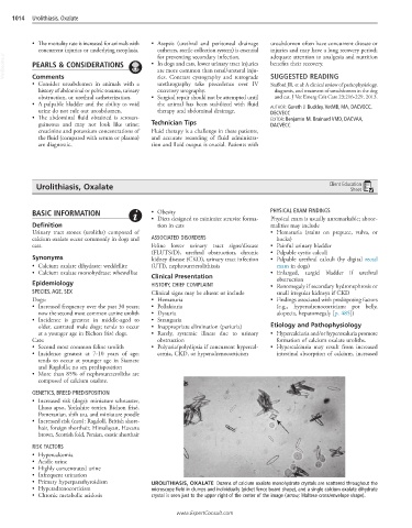
• Primary hyperparathyroidism UROLITHIASIS, OXALATE Dozens of calcium oxalate monohydrate crystals are scattered throughout the
• Hyperadrenocorticism microscope field in clumps and individually (picket fence board shape), and a single calcium oxalate dihydrate
• Chronic metabolic acidosis crystal is seen just to the upper right of the center of the image (arrow; Maltese cross/envelope shape).
www.ExpertConsult.com