
Page 212 - Cote clinical veterinary advisor dogs and cats 4th
P. 212
88 Atlantoaxial Instability
GENETICS, BREED PREDISPOSITION especially when the neck is ventroflexed. This ○ Ventrodorsal views
All toy/small breeds are predisposed: Yorkshire action causes the dens and cranial aspect of ■ If needed to determine dens hypoplasia
VetBooks.ir Pomeranians, Pekinese overrepresented; also in cord, resulting in contusive and compressive ■ For surgical planning
C2 to traumatize the cranial cervical spinal
or agenesis
terriers, miniature and toy poodles, Chihuahuas,
injury.
Cavalier King Charles spaniels
ASSOCIATED DISORDERS • Associated edema, inflammation +/− hemor- Advanced or Confirmatory Testing
• Plain radiographs usually sufficient to
rhage can further injure the spinal cord and
Other congenital anomalies: syringohydromy- extend into the caudal brainstem. diagnose the problem
elia, hydrocephalus, Chiari-like malformation, • Advanced imaging (MRI [p. 1132] or CT)
vertebral anomalies DIAGNOSIS often useful to rule out other regional dif-
Clinical Presentation Diagnostic Overview ferential diagnoses that can be coincident
with AAI, such as Chiari-like malformation
HISTORY, CHIEF COMPLAINT A tentative diagnosis is based on signalment, or syringomyelia.
• Variable onset of disease that can present history, and clinical signs. Confirmation requires • Note: If surgery is performed for AAI,
with acute, chronic, or intermittent signs. diagnostic imaging. nontitanium metallic implants prevent future
• Cervical pain is a consistent presenting MR/CT imaging of this anatomic region.
complaint. Ataxia, difficulty walking, or Differential Diagnosis
inability to ambulate can be the chief • Intervertebral disc disease (C2-C5) TREATMENT
complaint, depending on the severity of • Vertebral column trauma (C1-C5)
associated spinal cord injury. • Meningitis/myelitis: infectious or noninfec- Treatment Overview
• Seizure-like activity or “passing out” is tious (e.g., granulomatous meningoen- • AA joint reduction and stabilization
occasionally reported. cephalomyelitis) • Treatment of associated spinal cord injury
• Other congenital anomalies affecting the
PHYSICAL EXAM FINDINGS cervicomedullary junction Acute General Treatment
Range of neurologic deficits: • Vertebral column neoplasia (C1-C5) • Immobilization of the cervical spine
• Cervical pain alone • Discospondylitis (C2-C5) with the AA joint positioned in relative
• Cervical myelopathy (C1-C5) with varying extension
degrees of generalized ataxia and tetraparesis Initial Database ○ Soft, supportive, comfortable bandage
• Stiff, slightly extended head and neck • CBC/serum biochemical profile/urinalysis: extending from the temporomandibular
carriage generally unremarkable (pre-anesthesia) joint (TMJ) to the caudal cervical region.
• Tetraplegia and hypoventilation (rare) in • Plain radiography under general anesthesia A Styrofoam coffee cup, cut open from
severely affected animals (preferred) or heavy sedation because per- top to bottom with the bottom removed,
fectly positioned images are required to make can be gently wrapped around the neck,
Etiology and Pathophysiology the diagnosis. from just behind the ears (level of TMJ)
• Causes ○ Lateral vertebral column radiographic to the caudal cervical region and secured
○ Congenital malformation of the atlas findings with a strip of tape.
(C1) and/or axis (C2), with associated ■ Widened space between dorsal aspect • Cage confinement
malarticulation of C1 and C2 • Pain medication +/− antiinflammatory
○ Abnormal development of the dens of C2, ■ Dorsally displaced dens/cranial body medication
including agenesis, hypoplasia, and dorsal of C2 • Ventilation therapy (p. 1185) if respiratory
angulation and/or malformation; absence; ○ Oblique views compromise
or laxity of the ligaments that normally ■ If needed to visualize the dens (C2)
secure the dens onto the ventral aspect of ○ Stressed views Chronic Treatment
the C1 vertebral canal (transverse, dorsal ■ If needed to determine “instability” Surgical repair: AA joint reduction and
atlantoaxial, and/or alar ligaments). ■ Note: Use extreme caution whenever stabilization
○ Cervical trauma or hyperflexion injury moving the atlantoaxial (AA) joint • Indications
resulting in C2/dens fracture and/or liga- under anesthesia; use dorsiflexion rather ○ Most adult dogs; puppies with marked
mentous rupture than ventroflexion to demonstrate instability
• Any of the above can result in AAI, allowing instability to prevent further spinal ○ Any dog with recurrent neurologic signs
C2 to subluxate dorsally relative to C1, cord injury. ○ Dorsally angulated dens
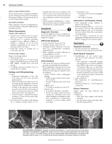
A B C D
ATLANTOAXIAL INSTABILITY Diagnosis of atlantoaxial instability in a 1-kg Yorkshire terrier. A, C, Plain lateral
radiographs and midsagittal T2-weighted MR image, neutral view, shows increased space between the dorsal lamina
of C1 and C2 (arrows). On the MRI, the dens can be seen compressing the spinal cord. B, D, With neck extension, the
subluxation reduces (arrow). C1-C2 transarticular pins provide stabilization and promote arthrodesis of the joint.
www.ExpertConsult.com