
Page 2204 - Cote clinical veterinary advisor dogs and cats 4th
P. 2204
1097.e2 Electromyography and Motor Nerve Conduction Velocity
(by continually redirecting the needle within
the muscle) and by increasing the number
of muscles sampled because denervation
VetBooks.ir potentials may be present in only a few
areas.
• A short burst of electrical activity that
occurs during needle placement is normal
due to temporary disruption of muscle fibers
(insertional activity). Other normal electrical
potentials called miniature endplate potentials
and endplate spikes may occur as a result of
spontaneous depolarization of part or all of
a myofiber.
• Normal healthy muscle is otherwise electri-
cally silent at rest.
• Other spontaneous electrical discharges in
resting muscle (i.e., anesthetized animal) A
are abnormal and indicate neuromuscular
disease.
• Abnormal EMG activity includes fibrillation
potentials, positive sharp waves, complex
repetitive discharges, or myotonic discharges.
In general, the type of waveform is not
specific for neuroanatomic location (nerve
vs. muscle) or pathophysiologic mechanism
of disease.
• Abnormal EMG activity confirms neuromus-
cular disease and helps localize which muscle
groups are affected; however, abnormal
EMG activity cannot differentiate between
myopathic and neuropathic disease.
• EMG changes due to denervation may
not be detected for 5-10 days after initial
injury.
• Severity of clinical signs does not always
correlate with severity of EMG changes. B
For the motor NCV procedure, begin with
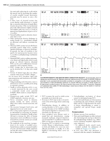
same preparation and anesthesia as for EMG, ELECTROMYOGRAPHY AND MOTOR NERVE CONDUCTION VELOCITY Electromyography: abnormal
above, and then spontaneous electrical activity. A, Fibrillation potentials, moderate density (100 mV/div, 10 msec/div). Fibrillation
• Using stimulating needle electrodes, a nerve is potentials may be associated with denervation secondary to axonopathy or with primary myopathy. Density
and consistency of observed fibrillation potentials are accurate reflections of severity of muscle involvement.
stimulated at various accessible points along B, Spontaneous electrical activity in the form of positive sharp waves (100 mV/div; 10 msec/div). (Reprinted
its anatomic pathway. with permission from Cuddon PA: Electrophysiology in neuromuscular disease. Vet Clin North Am Small Anim
• Needle or surface electrodes within or over Pract 32:31-62, 2002.)
a muscle innervated by the nerve record
a compound muscle action potential
(CMAP), which results in a simple biphasic
waveform.
• Measurements made include NCV and • NCV measures the speed at which action • Pathophysiologic mechanism of disease
CMAP amplitude, duration, and area. potentials travel down the axon. cannot be determined using NCV.
• Nerves commonly tested in domestic • Decreases in NCV may be due to demyelin- • Nerve and muscle biopsies are often necessary
animals include the sciatic, peroneal, ation or loss of the large, fastest conducting to determine the cause of disease, although
tibial, ulnar, radial, musculocutaneous, and axons. EMG and NCV help determine the nature
median. • Demyelinating diseases, in addition to of disease (demyelinating vs. axonal vs.
• The recurrent laryngeal, facial nerve, and causing decreases in NCV, cause a widen- myopathy) and optimal biopsy sample
trigeminal nerves are rarely tested. ing and distortion of the CMAP waveform site.
• NCV along various segments of axon may and temporal dispersion or distortion of the
be determined by measuring the distance waveform over the time axis. Postprocedure
between points of stimulation (ideally at least • Many peripheral nerve diseases result in • Monitor recovery from anesthesia.
100 mm to decrease the effect of human axonal loss and demyelination, causing • Nerve and muscle biopsies are often indicated
measurement error) and difference in latency decreased amplitude, temporal dispersion, after electrodiagnostic testing to determine
of waveforms (velocity [meters per second] and decreased NCV in addition to abnormal the pathophysiology of disease (e.g., inflam-
= Δ distance/Δ time). EMG activity due to denervation. matory, infectious, neoplastic, degenerative,
• The amplitude of the waveform is propor- • Measuring NCV is helpful in determining toxic, endocrine).
tional to the number of available axons. whether disease is demyelinating, axonal, ○ For additional details, consult recom-
Axonal disease results in a decreased number or both and whether the nerves are more mended methods for submission of
of axons and decreased CMAP amplitude affected proximally or distally. It is also samples in Shelton et al. (2002).
and may also cause secondary EMG changes helpful in determining which nerves are • In some cases, PEG tube placement is
due to denervation. affected. indicated while the animal is anesthetized.
www.ExpertConsult.com