
Page 2205 - Cote clinical veterinary advisor dogs and cats 4th
P. 2205
Electroretinogram 1097.e3
VetBooks.ir 1 1
3 3
Procedures and Techniques
5
5
A B
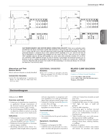
ELECTROMYOGRAPHY AND MOTOR NERVE CONDUCTION VELOCITY Motor nerve conduction studies
recorded from the plantar interosseous muscles after stimulation of the sciatic-tibial nerve at the hock, stifle, and
hip in a normal dog (A) and in a dog with generalized muscle disease (B) (mitochondrial myopathy). Note marked
generalized decrease in compound muscle action potential (CMAP) amplitudes (3.12, 3.37, and 4.51 mV) from all
sites of stimulation in the myopathic dog compared with the normal dog (23.02, 19.86, and 20.34 mV). Despite
CMAP amplitude reduction, the dog with myopathy has normal motor nerve conduction velocities (101 and 85 m/
sec). Generalized CMAP amplitude decrease can also be observed in prejunctional neuromuscular diseases such as
botulism, as well as in primary axonopathies. (Recording parameters: A, 5 mV/div and 2 msec/div; B, 1 mV/div and
2 msec/div.) (Reprinted with permission from Cuddon PA: Electrophysiology in neuromuscular disease. Vet Clin North
Am Small Anim Pract 32:31-62, 2002.)
Alternatives and Their ADDITIONAL SUGGESTED RELATED CLIENT EDUCATION
Relative Merits READING SHEET
Muscle and nerve biopsies as already described Shelton GD et al: Muscular dystrophies and other
inherited myopathies. Vet Clin North Am Small Consent to Perform General Anesthesia
SUGGESTED READING Anim Pract 32:103-124, 2002. AUTHOR: Greg Kilburn, DVM, DACVIM
Cuddon PA: Electrophysiology in neuromuscular EDITORS: Leah A. Cohn, DVM, PhD, DACVIM; Mark S.
disease. Vet Clin North Am Small Anim Pract Thompson, DVM, DABVP
32:31-62, 2002.
Electroretinogram
Difficulty level: ♦♦♦ rod-cone degeneration or progressive rod- cul-de-sac). Contact lens electrodes are used
cone dysplasias) at an early age well before most often.
Overview and Goal clinical signs and gross retinal changes occur • Reference and ground electrodes
• Evaluation of retinal function (quantitative • Evaluation of damage incurred by glaucoma • Standardized light source for light stimulus
or qualitative) through measurement of or toxic effects of drugs • “Safe” light, red light, or small red
the electrical response generated after the • Evaluation of the effects of optic nerve penlight
photoreceptors receive a light stimulus hypoplasia on retinal function • Timer for dark adaptation
• An electroretinogram (ERG) by itself does • Conducting agent to facilitate electrical
not determine whether an animal can see; the Contraindications contact between the contact lens electrode
retina is only one part of the visual system. • Panuveitis and the eye; methylcellulose (Gonak) or
• Corneal ulcer GenTeal eye gel
Indications • Any contraindication for general anesthesia • Table: if stainless steel, cover with noncon-
• Differentiation of causes of sudden blindness or sedation ducting material (rubber padding).
(e.g., sudden acquired retinal degeneration • Optional: head rest for elevation and posi-
syndrome [SARDS] and optic neuritis) Equipment, Anesthesia tioning of head; foam, rolled-up blankets,
• Evaluation of retinal function to determine Equipment: or sandbags
cataract surgery suitability • Computer and program for ERG Anesthesia or sedation:
• Identifying inherited retinal disease (i.e., • Gold-foil contact lens electrode or an atrau- • General anesthesia or sedation: some animals
progressive retinal atrophy such as progressive matic wire electrode (placed in conjunctival will tolerate the procedure with topical
www.ExpertConsult.com