
Page 2242 - Cote clinical veterinary advisor dogs and cats 4th
P. 2242
Foreign Body Removal, Esophageal (Endoscopic) 1115
Foreign Body Removal, Esophageal (Endoscopic) Client Education
Sheet
VetBooks.ir
• Mouth gag/speculum
Difficulty level: ♦♦
• Rigid tube (overtube) with smooth edges for • Antibiotic therapy if indicated by aspiration
pneumonia or esophageal perforation
Overview and Goal esophageal dilation (optional); typical external • Advise the owner of possible complications.
• In dogs, objects causing esophageal obstruc- diameters are 2 cm (cat, small dog), 3 cm ○ Emergency thoracotomy may be required
tion are mostly found in one of three loca- (medium-size dog), and 4 cm (large dog). if perforation occurs or removal by the oral
tions: at the thoracic inlet, at the level of • Flexible fiberoptic endoscope (or rigid route is impossible; possible gastrotomy if
the heart base, or caudal to the heart (most proctoscope) the foreign body has to be pushed through
common) (p. 351). • Endoscopic basket, grasping forceps (recom- into the stomach.
• Most foreign bodies obstructing the esopha- mended), polypectomy snare or endoscopic
gus can be removed without surgery. An biopsy forceps (second choice) Possible Complications and
attempt should be made to remove foreign • Water-soluble lubricating jelly Common Errors to Avoid Procedures and Techniques
bodies by esophageal endoscopy to avoid • Polyethylene catheter or feeding tube • Esophageal mucosal trauma (hemorrhage,
surgery and its complications (e.g., difficult (optional) erosion, ulceration) (p. 312)
access, limited healing ability, associated • Balloon catheter (optional) • Esophageal perforation, pyothorax (p. 857),
morbidity). • Suctioning apparatus pleuritis, mediastinitis
• If an object cannot be removed by the oral • Aspiration pneumonia
route, an attempt can be made to advance Anticipated Time • Tension pneumothorax (p. 797) (associated
it into the stomach, provided complications 20-90 minutes, depending on size of object and with esophageal insufflation)
such as esophageal perforation (from a ease with which it can be retrieved • Bradycardia due to vagal stimulation
sharp-edged foreign body, esophageal wall • Sepsis (p. 907) (due to aspiration or esopha-
devitalization, or overly aggressive forward Preparation: Important geal rupture)
pressure) are avoided. Objects passed into the Checkpoints • Esophageal stricture (p. 310) (clinically
stomach may be removed by gastrotomy or • Confirm location of foreign body, presence of manifests > 2 weeks postprocedure)
left to be digested (in the case of digestible aspiration pneumonia (p. 793) and evidence • Bronchoesophageal fistulation (rare)
objects). of esophageal perforation by performing
survey and contrast radiography using Procedure
Indications low-osmolality, nonionic contrast medium. • Classified as an emergency procedure
Foreign objects lodged in the esophagus (e.g., Radiographs should be performed immedi- • General anesthesia
bones, fishhooks, needles, toys, hairballs) ately before induction of general anesthesia • Place endotracheal tube and inflate cuff to
to confirm that the foreign body has not prevent aspiration of esophageal contents.
Contraindications spontaneously passed into the stomach. • Animal in sternal or left lateral recumbency
Esophageal perforation (p. 309) is an absolute • Endoscopic evaluation of location of foreign • Examine mouth and sublingual region for the
contraindication; thoracotomy is indicated in body and state of esophageal mucosa (p. presence of objects such as thread, needles,
these cases. 1098) or fishhooks.
• Ensure adequate patient hydration and • Suction esophagus to remove any liquid
Equipment, Anesthesia perfusion. contents and contrast medium.
• General anesthesia
• Cuffed endotracheal tube
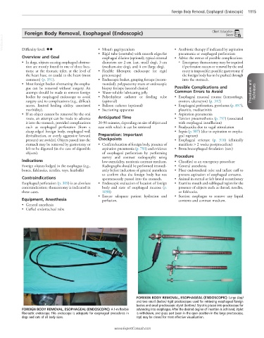
FOREIGN BODY REMOVAL, ESOPHAGEAL (ENDOSCOPIC) Large (top)
and two small (below) rigid proctoscopes used for retrieving esophageal foreign
bodies and small proctoscopic stylet (bottom). Stylet is placed into proctoscope for
FOREIGN BODY REMOVAL, ESOPHAGEAL (ENDOSCOPIC) A 1-m flexible advancing into esophagus. After the desired degree of insertion is achieved, stylet
fiberoptic endoscope. This endoscope is adequate for esophageal procedures in is withdrawn, and glass port (seen in the open position in the large proctoscope,
dogs and cats of all body sizes. top) may be closed for most effective visualization.
www.ExpertConsult.com