
Page 2284 - Cote clinical veterinary advisor dogs and cats 4th
P. 2284
Myelography 1134.e3
• Cisternal puncture (p. 1080) ○ Standard radiographic views (lateral, ven- ○ Compressing the jugular veins may
○ Commonly used for evaluating suspected ○ Optional views (lateral, neck flexed, neck increase CSF pressure, allowing CSF to
trodorsal, 45° oblique) are then obtained.
VetBooks.ir ○ Animal is placed in lateral recumbency extended, and traction views). CAUTION: ○ After CSF is obtained and no contraindica-
cervical spinal cord lesions
flow more readily.
with neck fully flexed at atlanto-occipital
neck-extended views may cause permanent
tions have been identified on CSF analysis,
joint.
particularly those with severe cervical
○ Insert needle on midline, with bevel damage to the spinal cord in some animals, attach tubing to the needle, and inject
contrast material slowly into subarachnoid
directed caudally, at the center of the IVDD. space.
triangle formed by the external occipital • Lumbar puncture ○ After contrast material is injected, the
protuberance and the wings of the atlas. ○ Commonly used for evaluating suspected needle is removed.
○ Advance needle slowly until ligamentum thoracolumbar lesions ○ Standard radiographic views (lateral, ven-
flavum and dorsal dura are punctured. ○ More technically difficult, fluoroscopy trodorsal, 45° oblique) are then obtained.
Because the puncturing may not be beneficial (p. 1080)
apparent, a good approach is to advance ○ Animal in lateral recumbency Postprocedure
1-2 mm at a time, removing the stylet and ○ Sixth lumbar spinous process is palpated. • The animal’s head should be elevated so
checking for CSF in the hub of the needle For anatomic localization of this spinous contrast material does not accumulate around Procedures and Techniques
each time before replacing the stylet and process (p. 1080) the brain.
advancing further. ○ With bevel directed cranially, introduce • The animal should be monitored for seizure
○ After the subarachnoid space is entered needle at 30°-60° angle at this site just to activity.
and CSF flows, obtain a CSF sample for the side of the spinous process. • If seizures are encountered, diazepam
immediate analysis. ○ Reposition needle until tip enters inter- 0.5-1 mg/kg IV can be administered.
○ If CSF analysis does not provide a diag- arcuate space between L5 and L6. It may • Many clinicians recommend keeping the
nosis, the procedure may be continued. be necessary to flex the spine, especially animal under general anesthesia for 30
○ Attach tubing to the needle and inject in older animals with degenerative bony minutes to 1 hour after myelography to
contrast material slowly. changes. decrease the incidence of seizures.
○ After contrast material is injected com- ○ The tail may twitch as the needle enters • Myelographic effect on CSF: increased cell
pletely, the needle is removed. the spinal cord. count (pleocytosis), increased percentage of
○ Elevate head before obtaining radiographs ○ Advance needle to canal floor.
to allow contrast material to flow cau- ○ Remove stylet and check for CSF; if no
dally from the atlanto-occipital site of CSF is visualized, withdraw needle slowly
injection. until flow is obtained.
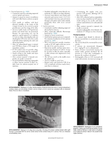
MYELOGRAPHY Myelogram of a dog, lateral projection. Moderate dorsal deviation of ventral subarachnoid
contrast column and attenuation of dorsal contrast column at L2-L3 (arrow) are consistent with an extradural
lesion. The L2-L3 disc is mineralized.
MYELOGRAPHY Myelogram of cat, ventrodorsal
projection. Lumbar and caudal thoracic portion. Normal
MYELOGRAPHY Myelogram of a dog, lateral projection. Thinning of the dorsal contrast column with a golf study. Incidental finding is a small amount of epidural
tee sign is associated with the ventral column dorsal to the vertebral body of C7 (arrow). This is an intradural accumulation of contrast material in the caudal lumbar
extramedullary lesion. region (bottom).
www.ExpertConsult.com