
Page 2322 - Cote clinical veterinary advisor dogs and cats 4th
P. 2322
1151.e8 Physical Rehabilitation
• Treatment times generally last 15-20 minutes Low-level laser therapy: • Changes in body composition
for 3-4 times per day. • A modality that uses the application of light ○ Evaluate patient on a scale of 1 to 9, in
VetBooks.ir • Water buoyancy decreases concussive forces • Clinically, this technique is employed to • Pain assessment analysis using a simple score
which 1 = emaciated and 9 = obese. A
energy to interact with and stimulate cells,
Aquatic therapy:
cell membranes, proteins, and DNA/RNA
body condition score of 4-5/9 is ideal.
and weight bearing on joints, and resistance
increases strength and helps maintain lean
proliferation and epithelialization and for
0 No evidence of pain during palpation of
body mass. accelerate wound healing by increased cell chart
• Underwater treadmill analgesic effects. joint
○ Allows active ROM similar to the • Treatment is often initiated within the first 48 1 Mild pain during palpation
patient’s normal gait while optimizing hours of postoperative procedures. Dosages 2 Moderate pain during palpation
the buoyancy, warmth, and resistance of used in dogs and cats for acute injuries are 3 Severe pain during palpation
2
water. typically less than 2 J/cm , whereas subacute 4 Patient does not permit clinician to palpate
2
○ Techniques can involve interval training injuries are typically 3-4 J/cm . Dogs with joint
(short durations of walking alternating osteoarthritis may be administered higher • Return to function
with periods of rest), continuous walking, doses of 8-10 J/cm . 2 ○ The desired level of function depends
or jogging. on the previous lifestyle of the patient
• Pool therapy: performed in a swimming pool Postprocedure (working dog vs. typical household pet).
or deep body of water Monitoring is vital to assess the patient’s • Quality of life assessment
○ Eliminates virtually all weight-bearing progress over time and to improve treatment • After a physical rehabilitation program is
forces protocols. Useful tools or assessment criteria complete, precaution is needed to avoid
○ Techniques include interval swimming for evaluating outcomes: overexertion and risk of relapse of clinical
(short durations of swimming, assisted or • Gait analysis signs. In many cases, a gradual increase in
on own, alternating with periods of rest), ○ Evaluate weight bearing at a stance; physical activity is necessary for optimal
free swimming (following the clinician or perform a lameness evaluation at a walk return to function.
owner across a specified distance), resisted and a trot.
swimming (patient is restrained by holding ○ Kinetic and kinematic (force plate and Alternatives and Their
the harness or life jacket), or swimming motion) gait analysis if available Relative Merits
against a current. • Goniometry Acupuncture (p. 1056): several studies have
Transcutaneous electrical nerve stimulation ○ Measure the available ROM of a joint and demonstrated enhanced postoperative analgesia
(TENS): compare with starting measurements. in dogs and improved neurologic function after
• With application of high-frequency current ○ Flex/extend joint until signs of discomfort spinal cord injury using acupuncture techniques.
directly to the skin by electrodes, the current are noted.
disrupts the normal sensory pathways for ○ Average of three measurements are taken. SUGGESTED READING
pain perception and provides analgesia. • Pedometers or accelerometers Millis DL, et al: Canine rehabilitation and physical
• Typically employed immediately postopera- ○ Evaluate trends in physical activity over therapy, ed 2, Philadelphia, 2014, Elsevier.
tively and during therapy to assist with pain time.
management • Assessment of muscle mass RELATED CLIENT EDUCATION
• Treatment duration and frequency vary, but ○ Record muscle girth using a measuring SHEETS
in general, low-intensity, short intervals are tape with spring tension device.
used during the acute stages (15 minutes; ○ For consistency, evaluate the hindlimb at How to Assist a Pet That Is Unable to Rise
1-2 times daily), and higher intensities and three quarters of the distance between the and Walk
longer intervals are used for chronic condi- greater trochanter to the patella. How to Perform Range-of-Motion Exercises
tions (up to 30 minutes; 2-3 times/week for ○ Evaluate the forelimb at three quarters of
5-6 weeks). the distance from the greater tubercle to AUTHOR: Allison Wara, DVM, DACVN
Neuromuscular electrical stimulation (NMES): the olecranon. EDITORS: Leah A. Cohn, DVM, PhD, DACVIM; Mark S.
• Application of low-level electrical current Thompson, DVM, DABVP
stimulates motor nerves and muscle fibers,
generating a muscle contraction.
• Can be used to combat muscle atrophy from
disuse and neurogenic causes and may assist
with muscle re-education, decreasing edema,
and providing analgesia
• Treatment duration and frequency are typi-
cally 15-20 minutes, 3-7 times per week.
Therapeutic ultrasound:
• Converts electricity to sound waves through
a transducer head, producing vibration in
the underlying tissue
• Stimulates fibroblast activity, increases circu-
lation and protein synthesis, and promotes
tissue healing.
• The intensity can be adjusted from 1-3 MHz.
As the frequency increases, penetration
decreases over an available range of 0.5-5 cm
tissue depth.
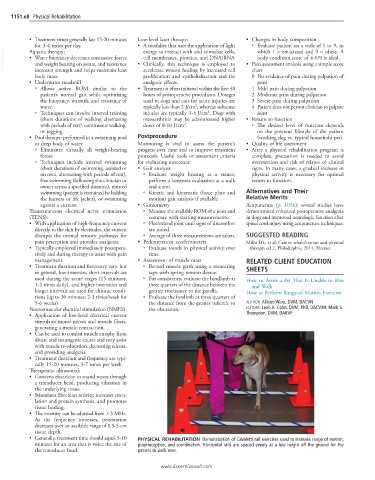
• Generally, treatment time should equal 5-10 PHYSICAL REHABILITATION Demonstration of Cavaletti rail exercises used to increase range of motion,
minutes for an area that is twice the size of proprioception, and coordination. Horizontal rails are spaced evenly at a low height off the ground for the
the transducer head. patient to walk over.
www.ExpertConsult.com