
Page 2319 - Cote clinical veterinary advisor dogs and cats 4th
P. 2319
Phlebotomy 1151.e5
• Ensure patient has appropriately been inversion 5-10 times to any tubes containing should fill with the appropriate volume of
prepared for desired testing (e.g., fasted; • A light wrap is placed if the patient is known • If the tube does not fill, redirect the needle
blood if the needle is in the jugular vein.
anticoagulant.
VetBooks.ir • Experienced restrainers reduce the stress Collecting blood with a Vacutainer system from to locate the vessel. Do not remove the
appropriate timing in regard to medications).
or suspected to be coagulopathic.
needle from the neck with the Vacutainer
to the patient and improve the chance for
successful venipuncture.
the jugular vein:
• Assemble the Vacutainer needle in the holder, tube attached because the tube will lose
vacuum.
Possible Complications and and have collection tubes nearby (see first ○ If it seems that the needle should be
Common Errors to Avoid Video). in the vein but there is no blood flow,
• Hematoma or bruising at collection site: • Patient is restrained in a sitting position; consider that the tube has lost vacuum
hold off afterward or apply wrap, especially sternal recumbency or standing positions are (try a new tube) or that the needle has
if problems with coagulation are recognized acceptable alternatives. gone all the way through and out of the
• Hemolyzed sample: minimized by jugular • An assistant positions the head and neck for vein (try backing up and finding the vein
venipuncture, “clean stick”, using a larger- access to the jugular vein by pointing the in more superficial tissue).
gauge needle, and allowing vacuum tubes nose upward at about a 45°-70° angle. • When ready to change tubes, release pressure
to fill without assistance • Wet the jugular furrow with 70% isopropyl on the vein. While steadying the Vacutainer Procedures and Techniques
• Underfilled collection tubes: results of certain alcohol to aid in visualization (clipping a holder and needle in place, remove the full
tests can be greatly affected by incomplete small patch is optional). tube. After the tube is out, rock to mix
filling (e.g., blue-top tubes used in coagula- • The phlebotomist occludes the jugular vein sample with anticoagulant.
tion assays). For small patients, consider use by applying pressure at the thoracic inlet with • Grab the next tube ready to be filled, reapply
of pediatric collection tubes. the nondominant hand. Laying the thumb pressure to the vein, and insert the tube onto
• Unable to obtain sample: restraint is key crossways just above the thoracic inlet is the inner needle in the Vacutainer assembly
to sample collection. If unable to obtain preferred to pushing the thumb into the as before.
sample after several tries, allow a colleague thoracic inlet. • Repeat for as many tubes as necessary.
to try. • The phlebotomist inserts the needle with the • After all tubes are filled, remove the last
bevel up through the skin and subcutaneous tube before removing the Vacutainer needle
Procedure tissue into the jugular vein; you will not see assembly.
• Select site: jugular vein, cephalic vein, medial a flash of blood (see second Video). • Apply light pressure to venipuncture site.
(cat) or lateral (dog) saphenous vein used • After the Vacutainer needle assembly is in
most often place, it need not be held. The phlebotomist Postprocedure
• Shaving is rarely necessary for collection of can let go of the assembly and pick up a • If there is reason to suspect continued bleed-
diagnostic blood samples, but shaving a very nearby collection tube or may have started ing, place a light wrap on the collection site
small patch can make it easier to see/feel with a tube in hand. for ≈15-30 minutes.
the vessel in some patients (e.g., those with • Push a Vacutainer collection tube onto the • Blood is best cleaned from the fur with water
thick, long coats) internal needle to start blood flow. The tube and then hydrogen peroxide (if needed).
Collecting blood with a syringe and needle
system from a peripheral vein:
• Patient positioning
○ Lateral recumbency for the lateral or
medial saphenous venipuncture
○ Sternal recumbency (or sitting) for cephalic
venipuncture
• Assistant holds the limb in position while
phlebotomist wets the fur with 70% isopro-
pyl alcohol (shaving first if appropriate).
• Assistant occludes the vessel to make the
vessel engorge distal to occlusion.
○ Alternatively, a tourniquet can be applied
to the limb.
• Phlebotomist holds the distal limb with the
nondominant hand with thumb parallel to
the vessel.
• Hold the assembled choice of needle and
syringe in the dominant hand, and insert
the needle through the skin into the vessel
with the needle bevel up.
• Apply small amounts of negative pressure
to the syringe to fill it without collapsing
the vessel.
• Fill syringe to desired amount, and notify
the assistant that you are finished so he or
she can stop occluding the vessel (or loosen A B
the tourniquet).
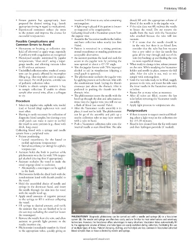
• Remove the needle from the vein, and allow PHLEBOTOMY Diagnostic phlebotomy can be carried out with a needle and syringe (A) or a Vacutainer
system (B). The needle and syringe are often less costly and are familiar to most veterinarians and veterinary
assistant to provide light pressure to the technicians. However, the Vacutainer offers advantages in many situations. Vacutainer tubes automatically fill
needle insertion site. to the appropriate volume of blood, and tube types are easily switched during collection, facilitating the use
• Phlebotomist immediately transfers the blood of multiple types of tubes. Platelet clumping, clotting, and hemolysis are less common in Vacutainer-collected
to the appropriate tubes, quickly giving an blood samples than in those collected by needle and syringe.
www.ExpertConsult.com