
Page 2332 - Cote clinical veterinary advisor dogs and cats 4th
P. 2332
Radiographic Interpretation, Abdomen 1155.e3
VetBooks.ir
Procedures and Techniques
RADIOGRAPHIC INTERPRETATION, ABDOMEN Pyometra. Lateral abdominal
radiograph of a 4-year-old, female Labrador retriever. Multiple, large, tubular,
soft-tissue–opacity structures (black V) are present, filling the caudal abdomen,
displacing the distal extremity of the spleen, and displacing multiple small-intestinal RADIOGRAPHIC INTERPRETATION, ABDOMEN Small intestinal obstruc-
loops cranially. The curved gas opacity of the stomach is indicative of gastric tion. Right lateral radiographs of an 11-year-old Staffordshire terrier cross with a
compression (arrows). Sublumbar lymphadenomegaly (asterisk) causes ventral history of vomiting for 3 days. Multiple, moderately dilated, gas- and fluid-filled
colonic displacement. Severe uterine dilation and medial iliac lymphadenomegaly small-intestinal loops (arrows) are seen in the middle and cranial abdomen. Within
were confirmed ultrasonographically. one of these loops is an irregularly margined mineral opacity (asterisk). A second,
nondilated population of small intestines (V) can be seen in the mid-abdomen.
The abdominal serosal detail is normal. These radiographic signs are indicative of
a complete small-intestinal obstruction. On ultrasound examination, the foreign
body had moved to the colon, and a piece of bone was passed in the feces after
conservative management.
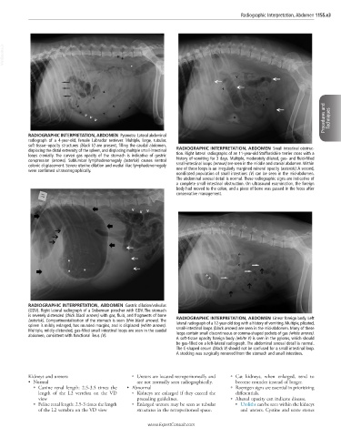
RADIOGRAPHIC INTERPRETATION, ABDOMEN Gastric dilation/volvulus
(GDV). Right lateral radiograph of a Doberman pinscher with GDV. The stomach
is severely distended (thick black arrows) with gas, fluid, and fragments of bone
(asterisk). Compartmentalization of the stomach is seen (thin black arrows). The RADIOGRAPHIC INTERPRETATION, ABDOMEN Linear foreign body. Left
spleen is mildly enlarged, has rounded margins, and is displaced (white arrows). lateral radiograph of a 12-year-old dog with a history of vomiting. Multiple, plicated,
Multiple, mildly distended, gas-filled small intestinal loops are seen in the caudal small-intestinal loops (black arrows) are seen in the mid-abdomen. Many of these
abdomen, consistent with functional ileus (V). loops contain small discontinuous or comma-shaped pockets of gas (white arrows).
A soft-tissue opacity foreign body (white V) is seen in the pylorus, which should
be gas-filled on a left-lateral radiograph. The abdominal serosal detail is normal.
The C-shaped cecum (black V) should not be confused for a small intestinal loop.
A stocking was surgically removed from the stomach and small intestines.
Kidneys and ureters: ○ Ureters are located retroperitoneally and ○ Cat kidneys, when enlarged, tend to
• Normal are not normally seen radiographically. become rounder instead of longer.
○ Canine renal length: 2.5-3.5 times the • Abnormal ○ Roentgen signs are essential in prioritizing
length of the L2 vertebra on the VD ○ Kidneys are enlarged if they exceed the differentials.
view preceding guidelines. ○ Altered opacity can indicate disease.
○ Feline renal length: 2.5-3 times the length ○ Enlarged ureters may be seen as tubular ■ Uroliths can be seen within the kidneys
of the L2 vertebra on the VD view structures in the retroperitoneal space. and ureters. Cystine and urate stones
www.ExpertConsult.com