
Page 2389 - Cote clinical veterinary advisor dogs and cats 4th
P. 2389
1182 Urinary Catheter Care, Indwelling
VetBooks.ir D
A B C
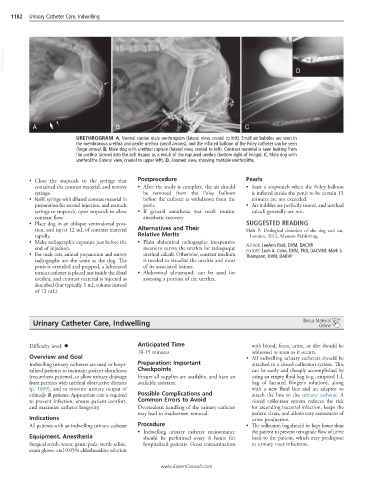
URETHROGRAM A, Normal canine male urethrogram (lateral view, cranial to left). Small air bubbles are seen in
the membranous urethra and penile urethra (small arrows), and the inflated balloon of the Foley catheter can be seen
(large arrow). B, Male dog with urethral rupture (lateral view, cranial to left). Contrast material is seen leaking from
the urethra (arrow) into the soft tissues as a result of the ruptured urethra (bottom right of image). C, Male dog with
urethroliths (lateral view, cranial to upper left). D, Zoomed view, showing multiple urethroliths.
• Close the stopcock to the syringe that Postprocedure Pearls
contained the contrast material, and remove • After the study is complete, the air should • Start a stopwatch when the Foley balloon
syringe. be removed from the Foley balloon is inflated inside the penis to be certain 15
• Refill syringe with diluted contrast material in before the catheter is withdrawn from the minutes are not exceeded.
preparation for second injection, and reattach penis. • Air bubbles are perfectly round, and urethral
syringe to stopcock; open stopcock to allow • If general anesthesia was used: routine calculi generally are not.
contrast flow. anesthetic recovery
• Place dog in an oblique ventrodorsal posi- SUGGESTED READING
tion, and inject 12 mL of contrast material Alternatives and Their Holt P: Urological disorders of the dog and cat,
rapidly. Relative Merits London, 2012, Manson Publishing.
• Make radiographic exposure just before the • Plain abdominal radiographs: inexpensive
end of injection. means to survey the urethra for radiopaque AUTHOR: LeeAnn Pack, DVM, DACVR
• For male cats, animal preparation and survey urethral calculi. Otherwise, contrast medium EDITORS: Leah A. Cohn, DVM, PhD, DACVIM; Mark S.
Thompson, DVM, DABVP
radiographs are the same as the dog. The is needed to visualize the urethra and most
penis is extruded and prepped, a lubricated of its associated lesions.
tomcat catheter is placed just inside the distal • Abdominal ultrasound: can be used for
urethra, and contrast material is injected as assessing a portion of the urethra.
described (but typically 3 mL volume instead
of 12 mL).
Urinary Catheter Care, Indwelling Bonus Material
Online
Anticipated Time
Difficulty level: ♦ with blood, feces, urine, or dirt should be
10-15 minutes addressed as soon as it occurs.
Overview and Goal • All indwelling urinary catheters should be
Indwelling urinary catheters are used in hospi- Preparation: Important attached to a closed collection system. This
talized patients to maintain patient cleanliness Checkpoints can be easily and cheaply accomplished by
(recumbent patients), to allow urinary drainage Ensure all supplies are available, and have an using an empty fluid bag (e.g., emptied 1-L
from patients with urethral obstructive diseases available assistant. bag of lactated Ringer’s solution), along
(p. 1009), and to monitor urinary output of with a new fluid line and an adaptor to
critically ill patients. Appropriate care is required Possible Complications and attach the line to the urinary catheter. A
to prevent infection, ensure patient comfort, Common Errors to Avoid closed collection system reduces the risk
and maximize catheter longevity. Overzealous handling of the urinary catheter for ascending bacterial infection, keeps the
may lead to inadvertent removal. patient clean, and allows easy assessment of
Indications urine production.
All patients with an indwelling urinary catheter Procedure • The collection bag should be kept lower than
• Indwelling urinary catheter maintenance the patient to prevent retrograde flow of urine
Equipment, Anesthesia should be performed every 8 hours for back to the patient, which may predispose
Surgical scrub, water, gauze pads, sterile saline, hospitalized patients. Gross contamination to urinary tract infections.
exam gloves, and 0.05% chlorhexidine solution
www.ExpertConsult.com