
Page 2496 - Cote clinical veterinary advisor dogs and cats 4th
P. 2496
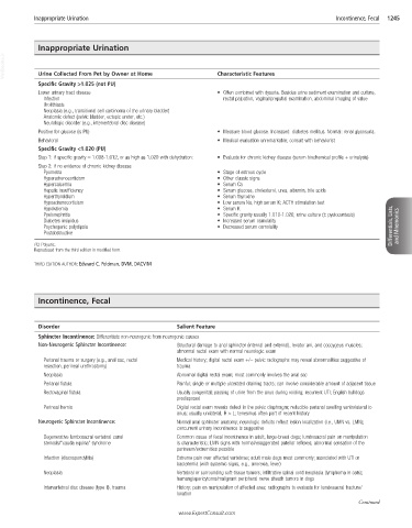
Inappropriate Urination                                                                   Incontinence, Fecal   1245


            Inappropriate Urination
  VetBooks.ir  Urine Collected From Pet by Owner at Home          Characteristic Features


            Specific Gravity >1.025 (not PU)
            Lower urinary tract disease                           •  Often combined with dysuria. Besides urine sediment examination and culture,
              Infection                                            rectal palpation, vaginal/preputial examination, abdominal imaging of value
              Urolithiasis
              Neoplasia (e.g., transitional cell carcinoma of the urinary bladder)
              Anatomic defect (pelvic bladder, ectopic ureter, etc.)
              Neurologic disorder (e.g., intervertebral disc disease)
            Positive for glucose (is PU)                          •  Measure blood glucose. Increased: diabetes mellitus. Normal: renal glycosuria.
            Behavioral                                            •  Medical evaluation unremarkable; consult with behaviorist
            Specific Gravity <1.020 (PU)
            Step 1: if specific gravity = 1.008-1.012, or as high as 1.020 with dehydration:  •  Evaluate for chronic kidney disease (serum biochemical profile + urinalysis)
            Step 2: if no evidence of chronic kidney disease:
              Pyometra                                            •  Stage of estrous cycle
              Hyperadrenocorticism                                •  Other classic signs
              Hypercalcemia                                       •  Serum Ca
              Hepatic insufficiency                               •  Serum glucose, cholesterol, urea, albumin, bile acids
              Hyperthyroidism                                     •  Serum thyroxine
              Hypoadrenocorticism                                 •  Low serum Na, high serum K; ACTH stimulation test
              Hypokalemia                                         •  Serum K
              Pyelonephritis                                      •  Specific gravity usually 1.010-1.020; urine culture (± pyelocentesis)
              Diabetes insipidus                                  •  Increased serum osmolality
              Psychogenic polydipsia                              •  Decreased serum osmolality                       Differentials, Lists,   and Mnemonics
              Postobstructive

           PU, Polyuric.
           Reproduced from the third edition in modified form.
           THIRD EDITION AUTHOR: Edward C. Feldman, DVM, DACVIM


            Incontinence, Fecal


            Disorder                                  Salient Feature
            Sphincter Incontinence: Differentiate non-neurogenic from neurogenic causes
            Non-Neurogenic Sphincter Incontinence:    Structural damage to anal sphincter (internal and external), levator ani, and coccygeus muscles;
                                                      abnormal rectal exam with normal neurologic exam
              Perianal trauma or surgery (e.g., anal sac, rectal   Medical history; digital rectal exam +/− pelvic radiographs may reveal abnormalities suggestive of
              resection, perineal urethrostomy)       trauma
              Neoplasia                               Abnormal digital rectal exam; most commonly involves the anal sac
              Perianal fistula                        Painful; single or multiple ulcerated draining tracts; can involve considerable amount of adjacent tissue
              Rectovaginal fistula                    Usually congenital; passing of urine from the anus during voiding; recurrent UTI; English bulldogs
                                                      predisposed
              Perineal hernia                         Digital rectal exam reveals defect in the pelvic diaphragm; reducible perianal swelling ventrolateral to
                                                      anus; usually unilateral, R > L; tenesmus often part of recent history
            Neurogenic Sphincter Incontinence:        Normal anal sphincter anatomy; neurologic deficits reflect lesion localization (i.e., UMN vs. LMN);
                                                      concurrent urinary incontinence is suggestive
              Degenerative lumbosacral vertebral canal   Common cause of fecal incontinence in adult, large-breed dogs; lumbosacral pain on manipulation
              stenosis/“cauda equina” syndrome        is characteristic; LMN signs with normal/exaggerated patellar reflexes; abnormal sensation of the
                                                      perineum/extremities possible
              Infection (discospondylitis)            Extreme pain over affected vertebrae; adult male dogs most commonly; associated with UTI or
                                                      bacteremia (with systemic signs, e.g., anorexia, fever)
              Neoplasia                               Vertebral or surrounding soft-tissue tumors; infiltrative spinal cord neoplasia (lymphoma in cats);
                                                      hemangiopericytoma/malignant peripheral nerve sheath tumors in dogs
              Intervertebral disc disease (type II), trauma  History; pain on manipulation of affected area; radiographs to evaluate for lumbosacral fracture/
                                                      luxation
                                                                                                            Continued
                                                      www.ExpertConsult.com