
Page 2493 - Cote clinical veterinary advisor dogs and cats 4th
P. 2493
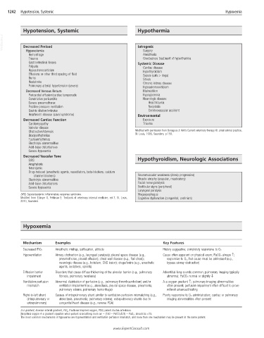
1242  Hypotension, Systemic                                                                          Hypoxemia


            Hypotension, Systemic                                Hypothermia
  VetBooks.ir  Decreased Preload                                 Iatrogenic


             Hypovolemia
                                                                   Anesthesia
              Hemorrhage                                           Surgery
              Trauma                                               Overzealous treatment of hyperthermia
              Gastrointestinal losses                            Systemic Disease
              Polyuria                                             Cardiac disease
              Hypoadrenocorticism                                  Hypothyroidism
              Effusions or other third spacing of fluid            Sepsis (cats > dogs)
              Burns                                                Shock
              Heatstroke                                           Chronic kidney disease
              Pulmonary arterial hypertension (severe)             Hypoadrenocorticism
             Decreased Venous Return                               Malnutrition
              Pericardial effusion/cardiac tamponade               Hypoglycemia
              Constrictive pericarditis                            Neurologic disease:
              Severe pneumothorax                                     Head trauma
              Positive-pressure ventilation                           Neoplasia
              Gastric dilation/volvulus                               Cerebrovascular accident
              Heartworm disease (caval syndrome)                 Environmental
            Decreased Cardiac Function                             Exposure
              Cardiomyopathy                                       Trauma
              Valvular disease
              Obstruction/stenosis                              Modified with permission from Bonagura J: Kirk’s Current veterinary therapy XII: small animal practice,
              Bradyarrhythmias                                  St. Louis, 1995, Saunders, p 159.
              Tachyarrhythmias
              Electrolyte abnormalities
              Acid-base disturbances
              Severe hypoxemia
            Decreased Vascular Tone
              SIRS                                               Hypothyroidism, Neurologic Associations
              Anaphylaxis
              Neurogenic
              Drug-induced (anesthetic agents, vasodilators, beta-blockers, calcium
                 channel blockers)                               Neuromuscular weakness (slowly progressive)
              Electrolyte abnormalities                          Muscle atrophy (scapular, masticatory)
              Acid-base disturbances                             Facial nerve paralysis
              Severe hypoxemia                                   Vestibular signs (peripheral)
                                                                 Laryngeal paralysis
           SIRS, Sepsis/systemic inflammatory response syndrome.  Megaesophagus
           Modified from Ettinger S, Feldman E: Textbook of veterinary internal medicine, ed 7, St. Louis,   Cognitive dysfunction (congenital; cretinism)
           2010, Saunders.


            Hypoxemia


            Mechanism      Examples                                          Key Features
                           Anesthetic mishap, suffocation, altitude
            Decreased FiO 2                                                  History suggestive; completely responsive to O 2
            Hypoventilation  Airway obstruction (e.g., laryngeal paralysis); pleural space disease (e.g.,   Cause often apparent on physical exam; PaCO 2 always ↑;
                             pneumothorax, pleural effusion); chest wall disease (e.g., flail chest);   responsive to O 2 , but cause must be addressed (e.g.,
                             neurologic disease (e.g., botulism, CNS lesion); drugs/toxins (e.g., anesthetic   bypass airway obstruction)
                             agents, sedatives, opioids)
            Diffusion barrier   Disorders that cause diffuse thickening of the alveolar barrier (e.g., pulmonary   Adventitial lung sounds common; pulmonary imaging typically
              impairment     fibrosis, pulmonary neoplasia)                   abnormal; PaCO 2 normal or slightly ↓
            Ventilation-perfusion   Abnormal distribution of perfusion (e.g., pulmonary thromboembolism) and/or   A-a oxygen gradient ↑; pulmonary imaging abnormalities
              mismatch       ventilation impairment (e.g., atelectasis, pleural space disease, pneumonia,   often present; perfusion impairment often difficult to prove
                             pulmonary edema, pulmonary hemorrhage)           without advanced testing
            Right-to-left shunt   Causes of intrapulmonary shunt similar to ventilation-perfusion mismatching (e.g.,   Poorly responsive to O 2 administration; cardiac or pulmonary
              (intrapulmonary or   atelectasis, pneumonia, pulmonary edema); extrapulmonary shunts due to   imaging abnormalities often present
              extrapulmonary)  congenital heart disease (e.g., reverse PDA)
           A-a gradient, alveolar-arterial gradient; FiO 2 , fractional inspired oxygen; PDA, patent ductus arteriosus.
           Simplified oxygen A-a gradient equation when patient is breathing room air = (150 – PaCO 2 /0.8) – PaO 2 ; should be <15.
           The most common mechanisms of hypoxemia are hypoventilation and ventilation-perfusion mismatch, and more than one mechanism may be present in the same patient.

                                                     www.ExpertConsult.com