
Page 2489 - Cote clinical veterinary advisor dogs and cats 4th
P. 2489
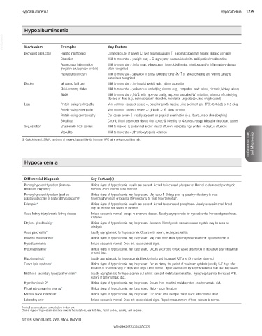
Hypoalbuminemia                                                                              Hypocalcemia   1239


            Hypoalbuminemia
  VetBooks.ir  Mechanism      Examples             Key Feature


            Decreased production  Hepatic insufficiency  Common cause of severe ↓; liver enzymes usually ↑; ± icterus; abnormal hepatic imaging common
                              Starvation           Mild to moderate ↓; weight loss; ± GI signs; may be associated with maldigestion/malabsorption
                              Acute phase inflammation   Mild to moderate ↓; inflammatory leukogram; hyperglobulinemia; infectious and/or inflammatory disease
                              (negative acute phase protein)  often recognized
                                                                                  +
                                                                                     +
                              Hypoadrenocorticism  Mild to moderate ↓; absence of stress leukogram; Na ↓K ↑ (if typical); waxing and waning GI signs
                                                   sometimes recognized
            Dilution          Iatrogenic fluid use  Mild to moderate ↓; in-hospital weight gain; history supportive
                              Fluid-retaining states  Mild to moderate ↓; evidence of underlying disease (e.g., congestive heart failure, cirrhosis, kidney failure)
                                                                                            +
                                                                 +
                              SIADH                Mild to moderate ↓; Na ↓ with hypo-osmolality; inappropriate urine Na  retention; evidence of underlying
                                                   disease or drug (e.g., nervous system disorders, neoplasia, lung disease, and drug induced)
            Loss              Protein-losing nephropathy  Very common cause of severe ↓; proteinuria with inactive urine sediment and UPC >0.4 (cat) or 0.5 (dog)
                              Protein-losing enteropathy  Very common cause of severe ↓; globulin ↓; GI signs common
                              Protein-losing dermatopathy  Can cause severe ↓; readily apparent on physical examination (e.g., burns, major skin sloughing)
                              Blood loss           Chronic blood loss more relevant than acute; GI bleeding or Ancylostoma spp infestation important causes
            Sequestration     Effusion into body cavities  Mild to marked ↓; abdominal and/or pleural effusion, especially high protein or chylous effusions
                                                                                                                      Differentials, Lists,   and Mnemonics
                              Vasculitis           Mild to moderate ↓; thrombocytopenia common
           GI, Gastrointestinal; SIADH, syndrome of inappropriate antidiuretic hormone; UPC, urine protein creatinine ratio.


            Hypocalcemia


            Differential Diagnosis         Key Feature(s)
            Primary hypoparathyroidism (immune-  Clinical signs of hypocalcemia usually are present. Normal to increased phosphorus. Normal to decreased parathyroid
            mediated, idiopathic)*         hormone (PTH). Normal renal function.
            Primary hypoparathyroidism (post-op   Clinical signs of hypocalcemia may be present. May occur 1-3 days post-op parathyroidectomy to treat
            parathyroidectomy or bilateral thyroidectomy)*  hyperparathyroidism or bilateral thyroidectomy to treat hyperthyroidism.
            Eclampsia*                     Clinical signs of hypocalcemia usually are present. Normal to decreased phosphorus. Usually occurs in small-breed
                                           dogs in the first few weeks of lactation.
            Acute kidney injury/chronic kidney disease  Ionized calcium is normal, except in advanced disease. Usually asymptomatic for hypocalcemia. Increased phosphorus.
                                           Azotemia.
            Ethylene glycol toxicity*      Clinical signs of hypocalcemia may be present. Azotemia. Monohydrate calcium oxalate crystals may be seen on
                                           urinalysis.
            Acute pancreatitis*            Usually asymptomatic for hypocalcemia. Occurs with severe, acute pancreatitis.
            Intestinal malabsorption*      Clinical signs of hypocalcemia may be present. May have concurrent hypomagnesemia and/or hypovitaminosis D.
            Hypoalbuminemia                Ionized calcium is normal. Does not cause clinical signs.
            Hypomagnesemia*                Clinical signs of hypocalcemia may be present. Occurs secondary to decreased absorption or increased gastrointestinal
                                           or renal loss.
            Rhabdomyolysis*                Usually asymptomatic for hypocalcemia. Myoglobinuria and increased AST and CK may be observed.
            Tumor lysis syndrome*          Clinical signs of hypocalcemia may be present. Occurs during the period of maximum cytolysis (usually 5-7 days after
                                           initiation of chemotherapy) in dogs with large tumor burden. Hyperkalemia and hyperphosphatemia may also be present.
            Nutritional secondary hyperparathyroidism*  Usually asymptomatic for hypocalcemia but exhibit pain and skeletal abnormalities. Hypophosphatemia. Increased PTH.
                                           History of a homemade diet.
            Hypovitaminosis D*             Clinical signs of hypocalcemia may be present. Occurs from intestinal malabsorption or a homemade diet.
            Phosphate-containing enemas*   Clinical signs of hypocalcemia may be present. History is confirmatory.
            Massive blood transfusion*     Clinical signs of hypocalcemia may be present. Can occur after multiple transfusions with citrated blood.
            Laboratory error               Ionized calcium is normal. Does not cause clinical signs. Repeat measurement of total calcium is normal.
           *Ionized serum calcium concentration is also low.
           Clinical signs of hypocalcemia include muscle fasciculations, ear twitching, facial rubbing, anxiety, and seizures.
           AUTHOR: Karen M. Tefft, DVM, MVSc, DACVIM
                                                      www.ExpertConsult.com