
Page 512 - Cote clinical veterinary advisor dogs and cats 4th
P. 512
230 Cutaneous Neoplasia
Cutaneous Neoplasia Bonus Material
Online
VetBooks.ir
BASIC INFORMATION
the pluripotential basal epithelial cells in the
epidermis and adnexa DIAGNOSIS
Definition • Hemangioma/hemangiosarcoma: benign or Diagnostic Overview
Benign or malignant tumor arising from cells malignant neoplasms arising from endothelial The only way to distinguish benign neoplasms
within the skin and adnexa. The most common cells of blood vessels from malignant neoplasms and non-neoplastic
cutaneous neoplasms in dogs are, in descend- • Histiocytoma: benign neoplasm that arises proliferative skin disease is histopathologic
ing order of frequency, lipoma, sebaceous from epidermal Langerhans cells examination of biopsy tissue samples. Malignant
gland hyperplasia/adenoma, mast cell tumor, • Infundibular keratinizing acanthoma: benign lesions typically show sudden onset, rapid
histiocytoma, and papilloma. In cats, basal neoplasms of hair follicle origin invasive growth, infiltration, +/− metastasis.
cell tumors are the most common, followed • Trichoepithelioma: benign neoplasms that
by squamous cell carcinoma, mast cell tumor, arise from keratinocytes that differenti- Differential Diagnosis
and fibrosarcoma. Squamous cell carcinoma ate toward all three segments of the hair • Bacterial and fungal granulomas
(p. 939) and mast cell tumor (canine [p. 634] follicle • Abscesses
and feline [p. 632]) are discussed separately. • Sebaceous gland hyperplasia/adenoma: • Sterile granuloma
epithelial growths arising from sebocytes • Pyogranuloma syndrome
Synonyms • Sterile nodular panniculitis
• Basal cell tumor: basal cell epithelioma HISTORY, CHIEF COMPLAINT
• Infundibular keratinizing acanthoma: Solitary to multiple cutaneous masses Initial Database
keratoacanthoma, intracutaneous cornifying • CBC, serum biochemistry profile, urinalysis
epithelioma PHYSICAL EXAM FINDINGS (if indicated): assess systemic abnormalities
• Basal cell tumor: solitary, well-circumscribed, • Cytologic exam (fine-needle aspiration):
Epidemiology firm to cystic, alopecic, commonly ulcer- ○ Basal cell tumor: small, round to cuboidal
SPECIES, AGE, SEX ated, often pigmented mass, typically epithelial cells arranged in groups or
• Dogs: 30% of all tumors arise within the located on the head, neck, shoulders, ribbons. Basal cell carcinomas are difficult
skin. or thorax. In cats, malignant lesions to differentiate cytologically from benign
• Cats: 20% of all tumors arise within the also can occur on the nasal planum and lesions.
skin. eyelids. ○ Histiocytoma: sheets of round cells with a
• Median age for cutaneous neoplasia is 10.5 • Cutaneous hemangioma/hemangiosarcoma: pale blue cytoplasm and variable sizes and
years for dogs and 12 years for cats. dermal or subcutaneous, solitary or multiple, shapes of the nuclei; variable numbers of
• Predisposition for histiocytoma in young oval masses or red to dark red plaques, usually neutrophils and lymphocytes, depending
dogs located along the limbs and ventral abdomen. on stage of growth and involution
In cats, bluish to reddish black nodules to ○ Sebaceous gland hyperplasia: clusters of
GENETICS, BREED PREDISPOSITION plaques lipid sebocytes
• Canine breeds with highest incidence of skin • Histiocytoma: solitary, well-circumscribed, • Histopathologic exam (biopsy)
tumors include the boxer, Scottish terrier, firm, erythematous, intradermal nodule
bullmastiff, Weimaraner, Kerry blue terrier, found most frequently on the head, limbs,
and Norwegian elkhound. and thorax. Fast growing but benign.
• Shar-peis tend to develop mast cell tumors Occasionally observed as multiple cutaneous
at a younger age (mean, 4 years). nodules or plaques
• Feline breeds with highest incidence are • Infundibular keratinizing acanthoma:
Siamese and Persian. most commonly found on the back, neck,
• Infundibular keratinizing acanthoma: gen- thorax, and limbs. Well-circumscribed
eralized form may have a hereditary basis in dermal or subcutaneous masses with a
Norwegian elkhound and keeshond. German pore opening to the skin surface; pore
shepherds and Old English sheepdogs also usually consists of a keratin plug; not
develop the generalized form. metastatic
• Trichoepithelioma: usually solitary, solid or
RISK FACTORS cystic, elevated, round, and well circum-
• Basal cell carcinoma: a strong correlation in scribed; frequently becomes ulcerated and
humans exists between exposure to ultraviolet alopecic
light and tumor development. This associa- • Sebaceous gland tumors: solitary or multiple,
tion has not been established in dogs and raised, wartlike to smooth, may ulcerate;
cats. most commonly found on limbs, trunk,
• Cutaneous hemangioma/hemangiosarcoma: eyelids, head
short-coated dogs with nonpigmented skin
in sun-exposed areas such as the glabrous Etiology and Pathophysiology
(hairless) skin of the ventral abdomen and • Neoplastic transformation relies on changes
white cats are at higher risk. in specific growth-regulating genes.
• Principal growth-regulating genes include
Clinical Presentation ○ Oncogenes that code for proteins that
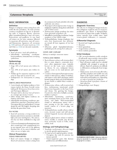
DISEASE FORMS/SUBTYPES increase growth CUTANEOUS NEOPLASIA Sebaceous adenoma
• Basal cell tumors: benign or malignant ○ Tumor-suppressor genes that decrease along the dorsal tail of a 7-year-old Labrador retriever.
(basal cell carcinoma) tumors arising from proliferation and differentiation (Copyright Dr. Ed Jazic.)
www.ExpertConsult.com